Chlorine »
PDB 4o0c-4oae »
4o9s »
Chlorine in PDB 4o9s: Crystal Structure of Retinol-Binding Protein 4 (RBP4)in Complex with A Non-Retinoid Ligand
Protein crystallography data
The structure of Crystal Structure of Retinol-Binding Protein 4 (RBP4)in Complex with A Non-Retinoid Ligand, PDB code: 4o9s
was solved by
Z.Wang,
S.Johnstone,
N.P.Walker,
with X-Ray Crystallography technique. A brief refinement statistics is given in the table below:
| Resolution Low / High (Å) | 30.00 / 2.30 |
| Space group | P 65 |
| Cell size a, b, c (Å), α, β, γ (°) | 85.737, 85.737, 118.806, 90.00, 90.00, 120.00 |
| R / Rfree (%) | 23.1 / 26.1 |
Other elements in 4o9s:
The structure of Crystal Structure of Retinol-Binding Protein 4 (RBP4)in Complex with A Non-Retinoid Ligand also contains other interesting chemical elements:
| Fluorine | (F) | 6 atoms |
Chlorine Binding Sites:
The binding sites of Chlorine atom in the Crystal Structure of Retinol-Binding Protein 4 (RBP4)in Complex with A Non-Retinoid Ligand
(pdb code 4o9s). This binding sites where shown within
5.0 Angstroms radius around Chlorine atom.
In total 3 binding sites of Chlorine where determined in the Crystal Structure of Retinol-Binding Protein 4 (RBP4)in Complex with A Non-Retinoid Ligand, PDB code: 4o9s:
Jump to Chlorine binding site number: 1; 2; 3;
In total 3 binding sites of Chlorine where determined in the Crystal Structure of Retinol-Binding Protein 4 (RBP4)in Complex with A Non-Retinoid Ligand, PDB code: 4o9s:
Jump to Chlorine binding site number: 1; 2; 3;
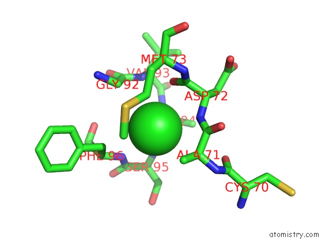
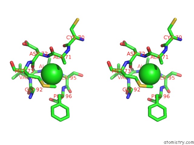
Chlorine binding site 1 out of 3 in 4o9s
Go back to
Chlorine binding site 1 out
of 3 in the Crystal Structure of Retinol-Binding Protein 4 (RBP4)in Complex with A Non-Retinoid Ligand
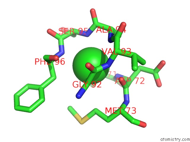
Mono view
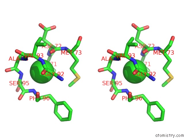
Stereo pair view
Mono view
Stereo pair view
A full contact list of Chlorine with other atoms in the Cl binding
site number 1 of Crystal Structure of Retinol-Binding Protein 4 (RBP4)in Complex with A Non-Retinoid Ligand within 5.0Å range:
|
Chlorine binding site 2 out of 3 in 4o9s
Go back to
Chlorine binding site 2 out
of 3 in the Crystal Structure of Retinol-Binding Protein 4 (RBP4)in Complex with A Non-Retinoid Ligand
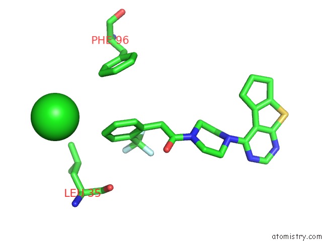
Mono view
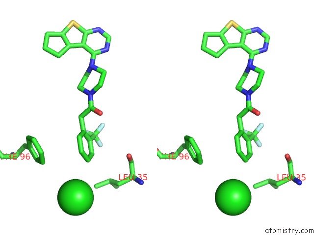
Stereo pair view
Mono view
Stereo pair view
A full contact list of Chlorine with other atoms in the Cl binding
site number 2 of Crystal Structure of Retinol-Binding Protein 4 (RBP4)in Complex with A Non-Retinoid Ligand within 5.0Å range:
|
Chlorine binding site 3 out of 3 in 4o9s
Go back to
Chlorine binding site 3 out
of 3 in the Crystal Structure of Retinol-Binding Protein 4 (RBP4)in Complex with A Non-Retinoid Ligand
Mono view
Stereo pair view
Mono view
Stereo pair view
A full contact list of Chlorine with other atoms in the Cl binding
site number 3 of Crystal Structure of Retinol-Binding Protein 4 (RBP4)in Complex with A Non-Retinoid Ligand within 5.0Å range:
|
Reference:
Y.Wang,
R.Connors,
P.Fan,
X.Wang,
Z.Wang,
J.Liu,
F.Kayser,
J.C.Medina,
S.Johnstone,
H.Xu,
S.Thibault,
N.Walker,
M.Conn,
Y.Zhang,
Q.Liu,
M.P.Grillo,
A.Motani,
P.Coward,
Z.Wang.
Structure-Assisted Discovery of the First Non-Retinoid Ligands For Retinol-Binding Protein 4. Bioorg.Med.Chem.Lett. V. 24 2885 2014.
ISSN: ISSN 0960-894X
PubMed: 24835984
DOI: 10.1016/J.BMCL.2014.04.089
Page generated: Fri Jul 11 19:55:08 2025
ISSN: ISSN 0960-894X
PubMed: 24835984
DOI: 10.1016/J.BMCL.2014.04.089
Last articles
Gd in 4AHXGd in 3ZTY
Gd in 4AHY
Gd in 3ZXS
Gd in 3VDZ
Gd in 3VX0
Gd in 3TXM
Gd in 3Q4I
Gd in 2YEU
Gd in 3H9V