Chlorine »
PDB 4ogv-4otj »
4ohs »
Chlorine in PDB 4ohs: The Structure of A Far-Red Fluorescent Protein, AQ143
Protein crystallography data
The structure of The Structure of A Far-Red Fluorescent Protein, AQ143, PDB code: 4ohs
was solved by
T.M.Wannier,
S.L.Mayo,
with X-Ray Crystallography technique. A brief refinement statistics is given in the table below:
| Resolution Low / High (Å) | 37.33 / 2.19 |
| Space group | P 1 |
| Cell size a, b, c (Å), α, β, γ (°) | 50.967, 68.114, 132.788, 98.38, 90.74, 110.47 |
| R / Rfree (%) | 19 / 22.1 |
Chlorine Binding Sites:
The binding sites of Chlorine atom in the The Structure of A Far-Red Fluorescent Protein, AQ143
(pdb code 4ohs). This binding sites where shown within
5.0 Angstroms radius around Chlorine atom.
In total 4 binding sites of Chlorine where determined in the The Structure of A Far-Red Fluorescent Protein, AQ143, PDB code: 4ohs:
Jump to Chlorine binding site number: 1; 2; 3; 4;
In total 4 binding sites of Chlorine where determined in the The Structure of A Far-Red Fluorescent Protein, AQ143, PDB code: 4ohs:
Jump to Chlorine binding site number: 1; 2; 3; 4;
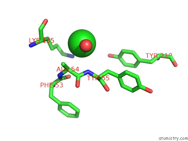
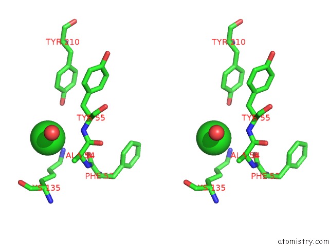
Chlorine binding site 1 out of 4 in 4ohs
Go back to
Chlorine binding site 1 out
of 4 in the The Structure of A Far-Red Fluorescent Protein, AQ143
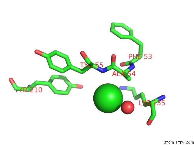
Mono view
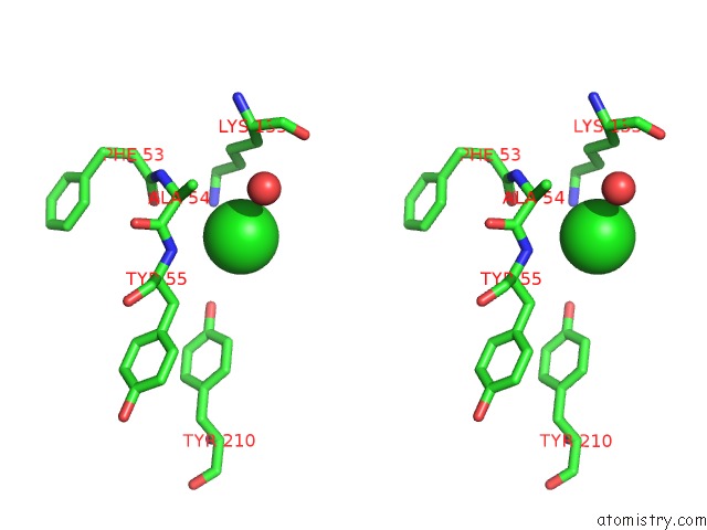
Stereo pair view
Mono view
Stereo pair view
|
|
A full contact list of Chlorine with other atoms in the Cl binding
site number 1 of The Structure of A Far-Red Fluorescent Protein, AQ143 within 5.0Å range:
|
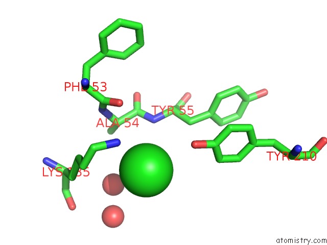
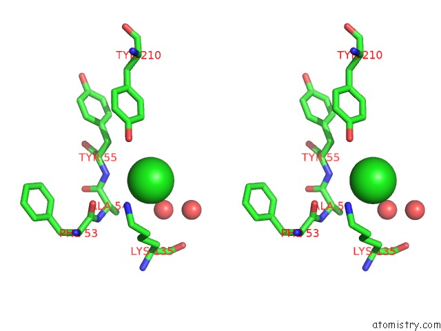
Chlorine binding site 2 out of 4 in 4ohs
Go back to
Chlorine binding site 2 out
of 4 in the The Structure of A Far-Red Fluorescent Protein, AQ143
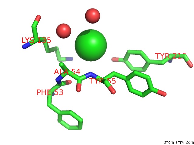
Mono view
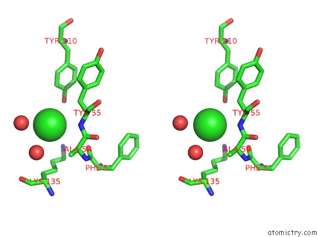
Stereo pair view
Mono view
Stereo pair view
|
|
A full contact list of Chlorine with other atoms in the Cl binding
site number 2 of The Structure of A Far-Red Fluorescent Protein, AQ143 within 5.0Å range:
|
Chlorine binding site 3 out of 4 in 4ohs
Go back to
Chlorine binding site 3 out
of 4 in the The Structure of A Far-Red Fluorescent Protein, AQ143
Mono view
Stereo pair view
Mono view
Stereo pair view
|
|
A full contact list of Chlorine with other atoms in the Cl binding
site number 3 of The Structure of A Far-Red Fluorescent Protein, AQ143 within 5.0Å range:
|
Chlorine binding site 4 out of 4 in 4ohs
Go back to
Chlorine binding site 4 out
of 4 in the The Structure of A Far-Red Fluorescent Protein, AQ143
Mono view
Stereo pair view
Mono view
Stereo pair view
|
|
A full contact list of Chlorine with other atoms in the Cl binding
site number 4 of The Structure of A Far-Red Fluorescent Protein, AQ143 within 5.0Å range:
|
Reference:
T.M.Wannier,
S.L.Mayo.
The Structure of A Far-Red Fluorescent Protein, AQ143, Shows Evidence in Support of Reported Red-Shifting Chromophore Interactions. Protein Sci. V. 23 1148 2014.
ISSN: ISSN 0961-8368
PubMed: 24888769
DOI: 10.1002/PRO.2498
Page generated: Fri Jul 11 20:02:57 2025
ISSN: ISSN 0961-8368
PubMed: 24888769
DOI: 10.1002/PRO.2498
Last articles
Mg in 6QJ6Mg in 6QJW
Mg in 6QJA
Mg in 6QIU
Mg in 6QIN
Mg in 6QGU
Mg in 6QII
Mg in 6QIE
Mg in 6QID
Mg in 6QEM