Chlorine »
PDB 4pk6-4pws »
4pm4 »
Chlorine in PDB 4pm4: Structure of A Putative Periplasmic Iron Siderophore Binding Protein (RV0265C) From Mycobacterium Tuberculosis H37RV
Protein crystallography data
The structure of Structure of A Putative Periplasmic Iron Siderophore Binding Protein (RV0265C) From Mycobacterium Tuberculosis H37RV, PDB code: 4pm4
was solved by
M.A.Arbing,
S.Chan,
N.Tran,
E.Kuo,
J.Lu,
L.R.Harris,
T.T.Zhou,
D.Eisenberg,
Tb Structural Genomics Consortium (Tbsgc),
with X-Ray Crystallography technique. A brief refinement statistics is given in the table below:
| Resolution Low / High (Å) | 29.20 / 2.20 |
| Space group | P 1 21 1 |
| Cell size a, b, c (Å), α, β, γ (°) | 69.551, 65.434, 72.330, 90.00, 116.41, 90.00 |
| R / Rfree (%) | 20.3 / 25.2 |
Chlorine Binding Sites:
The binding sites of Chlorine atom in the Structure of A Putative Periplasmic Iron Siderophore Binding Protein (RV0265C) From Mycobacterium Tuberculosis H37RV
(pdb code 4pm4). This binding sites where shown within
5.0 Angstroms radius around Chlorine atom.
In total 3 binding sites of Chlorine where determined in the Structure of A Putative Periplasmic Iron Siderophore Binding Protein (RV0265C) From Mycobacterium Tuberculosis H37RV, PDB code: 4pm4:
Jump to Chlorine binding site number: 1; 2; 3;
In total 3 binding sites of Chlorine where determined in the Structure of A Putative Periplasmic Iron Siderophore Binding Protein (RV0265C) From Mycobacterium Tuberculosis H37RV, PDB code: 4pm4:
Jump to Chlorine binding site number: 1; 2; 3;
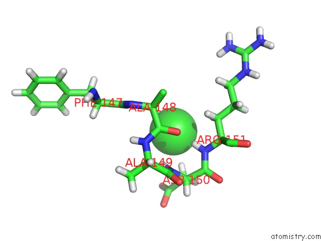
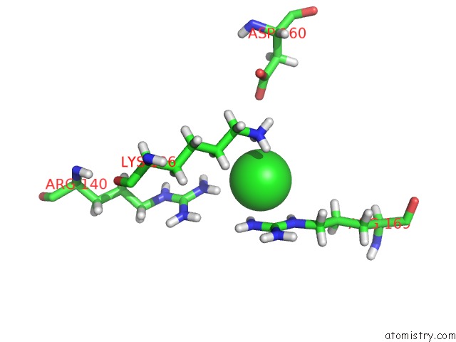
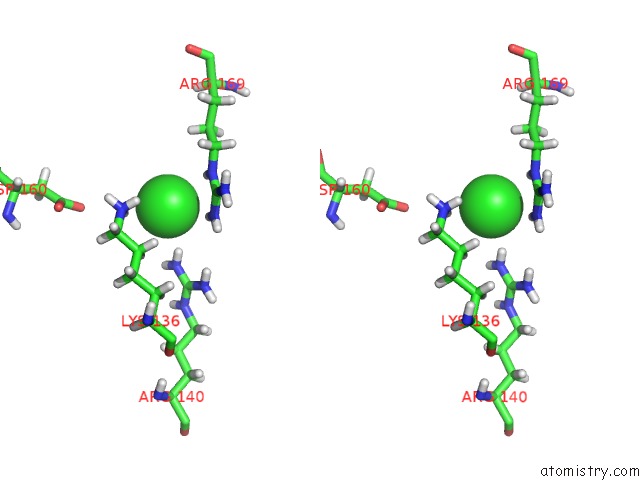
Chlorine binding site 1 out of 3 in 4pm4
Go back to
Chlorine binding site 1 out
of 3 in the Structure of A Putative Periplasmic Iron Siderophore Binding Protein (RV0265C) From Mycobacterium Tuberculosis H37RV
Mono view
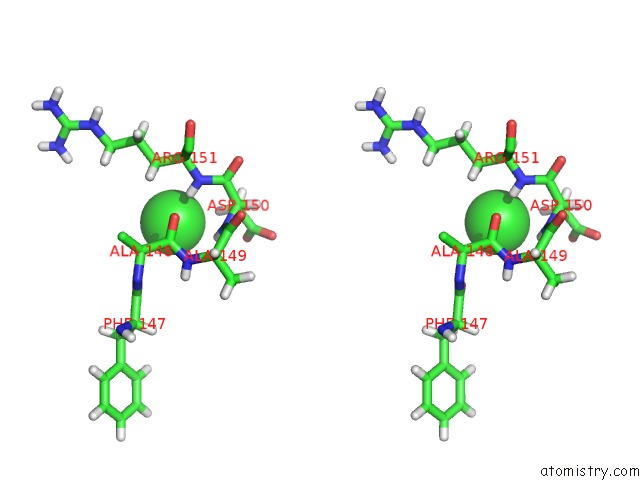
Stereo pair view
Mono view
Stereo pair view
A full contact list of Chlorine with other atoms in the Cl binding
site number 1 of Structure of A Putative Periplasmic Iron Siderophore Binding Protein (RV0265C) From Mycobacterium Tuberculosis H37RV within 5.0Å range:
|
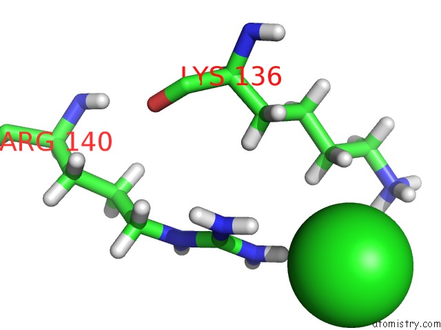
Chlorine binding site 2 out of 3 in 4pm4
Go back to
Chlorine binding site 2 out
of 3 in the Structure of A Putative Periplasmic Iron Siderophore Binding Protein (RV0265C) From Mycobacterium Tuberculosis H37RV
Mono view
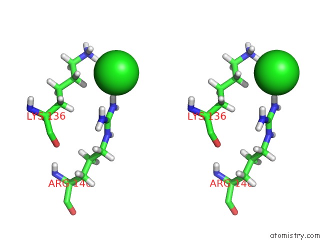
Stereo pair view
Mono view
Stereo pair view
A full contact list of Chlorine with other atoms in the Cl binding
site number 2 of Structure of A Putative Periplasmic Iron Siderophore Binding Protein (RV0265C) From Mycobacterium Tuberculosis H37RV within 5.0Å range:
|
Chlorine binding site 3 out of 3 in 4pm4
Go back to
Chlorine binding site 3 out
of 3 in the Structure of A Putative Periplasmic Iron Siderophore Binding Protein (RV0265C) From Mycobacterium Tuberculosis H37RV
Mono view
Stereo pair view
Mono view
Stereo pair view
A full contact list of Chlorine with other atoms in the Cl binding
site number 3 of Structure of A Putative Periplasmic Iron Siderophore Binding Protein (RV0265C) From Mycobacterium Tuberculosis H37RV within 5.0Å range:
|
Reference:
M.A.Arbing,
S.Chan,
N.Tran,
E.Kuo,
J.Lu,
L.R.Harris,
T.T.Zhou,
D.Eisenberg.
Structure of A Putative Periplasmic Iron Siderophore Binding Protein (RV0265C) From Mycobacterium Tuberculosis H37RV To Be Published.
Page generated: Fri Jul 11 20:30:34 2025
Last articles
Mg in 5BJOMg in 5B8F
Mg in 5B6A
Mg in 5B4L
Mg in 5B4K
Mg in 5B3S
Mg in 5B48
Mg in 5B47
Mg in 5B46
Mg in 5B30