Chlorine »
PDB 4wcv-4wkq »
4wji »
Chlorine in PDB 4wji: Crystal Structure of Cyclohexadienyl Dehydrogenase From Sinorhizobium Meliloti in Complex with Nadp and Tyrosine
Protein crystallography data
The structure of Crystal Structure of Cyclohexadienyl Dehydrogenase From Sinorhizobium Meliloti in Complex with Nadp and Tyrosine, PDB code: 4wji
was solved by
I.G.Shabalin,
D.R.Cooper,
J.Hou,
M.D.Zimmerman,
M.Stead,
B.S.Hillerich,
M.Ahmed,
J.Hammonds,
J.Bonanno,
R.Seidel,
S.C.Almo,
W.Minor,
New Yorkstructural Genomics Research Consortium (Nysgrc),
with X-Ray Crystallography technique. A brief refinement statistics is given in the table below:
| Resolution Low / High (Å) | 50.00 / 1.40 |
| Space group | C 1 2 1 |
| Cell size a, b, c (Å), α, β, γ (°) | 76.409, 68.891, 51.097, 90.00, 94.34, 90.00 |
| R / Rfree (%) | 11.5 / 15.9 |
Other elements in 4wji:
The structure of Crystal Structure of Cyclohexadienyl Dehydrogenase From Sinorhizobium Meliloti in Complex with Nadp and Tyrosine also contains other interesting chemical elements:
| Magnesium | (Mg) | 4 atoms |
Chlorine Binding Sites:
The binding sites of Chlorine atom in the Crystal Structure of Cyclohexadienyl Dehydrogenase From Sinorhizobium Meliloti in Complex with Nadp and Tyrosine
(pdb code 4wji). This binding sites where shown within
5.0 Angstroms radius around Chlorine atom.
In total only one binding site of Chlorine was determined in the Crystal Structure of Cyclohexadienyl Dehydrogenase From Sinorhizobium Meliloti in Complex with Nadp and Tyrosine, PDB code: 4wji:
In total only one binding site of Chlorine was determined in the Crystal Structure of Cyclohexadienyl Dehydrogenase From Sinorhizobium Meliloti in Complex with Nadp and Tyrosine, PDB code: 4wji:
Chlorine binding site 1 out of 1 in 4wji
Go back to
Chlorine binding site 1 out
of 1 in the Crystal Structure of Cyclohexadienyl Dehydrogenase From Sinorhizobium Meliloti in Complex with Nadp and Tyrosine
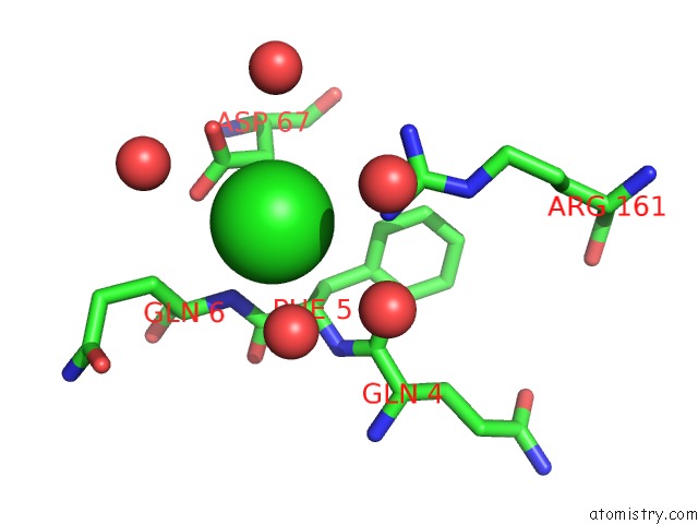
Mono view
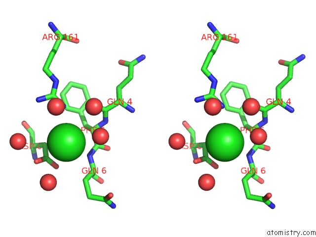
Stereo pair view
Mono view
Stereo pair view
A full contact list of Chlorine with other atoms in the Cl binding
site number 1 of Crystal Structure of Cyclohexadienyl Dehydrogenase From Sinorhizobium Meliloti in Complex with Nadp and Tyrosine within 5.0Å range:
|
Reference:
I.G.Shabalin,
D.R.Cooper,
J.Hou,
M.D.Zimmerman,
W.Minor.
Crystal Structure of Cyclohexadienyl Dehydrogenase From Sinorhizobium Meliloti in Complex with Nadp To Be Published.
Page generated: Fri Jul 11 22:32:40 2025
Last articles
Mg in 2WSBMg in 2WPD
Mg in 2WOQ
Mg in 2WOJ
Mg in 2WQS
Mg in 2WQN
Mg in 2WOG
Mg in 2WNL
Mg in 2WLL
Mg in 2WNH