Chlorine »
PDB 4wxp-4x97 »
4x1x »
Chlorine in PDB 4x1x: Crystal Structure of Rhdvb P Domain in Complex with Lewis Y
Protein crystallography data
The structure of Crystal Structure of Rhdvb P Domain in Complex with Lewis Y, PDB code: 4x1x
was solved by
M.M.Leuthold,
G.S.Hansman,
with X-Ray Crystallography technique. A brief refinement statistics is given in the table below:
| Resolution Low / High (Å) | 48.22 / 1.60 |
| Space group | P 1 21 1 |
| Cell size a, b, c (Å), α, β, γ (°) | 59.440, 84.150, 62.660, 90.00, 110.11, 90.00 |
| R / Rfree (%) | 15.5 / 18.5 |
Chlorine Binding Sites:
The binding sites of Chlorine atom in the Crystal Structure of Rhdvb P Domain in Complex with Lewis Y
(pdb code 4x1x). This binding sites where shown within
5.0 Angstroms radius around Chlorine atom.
In total 3 binding sites of Chlorine where determined in the Crystal Structure of Rhdvb P Domain in Complex with Lewis Y, PDB code: 4x1x:
Jump to Chlorine binding site number: 1; 2; 3;
In total 3 binding sites of Chlorine where determined in the Crystal Structure of Rhdvb P Domain in Complex with Lewis Y, PDB code: 4x1x:
Jump to Chlorine binding site number: 1; 2; 3;
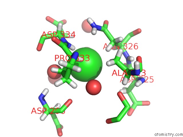
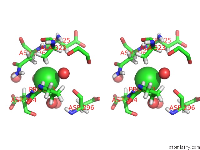
Chlorine binding site 1 out of 3 in 4x1x
Go back to
Chlorine binding site 1 out
of 3 in the Crystal Structure of Rhdvb P Domain in Complex with Lewis Y
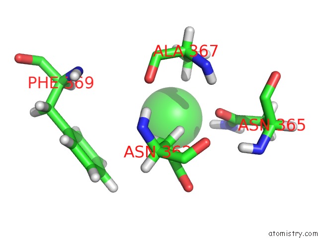
Mono view
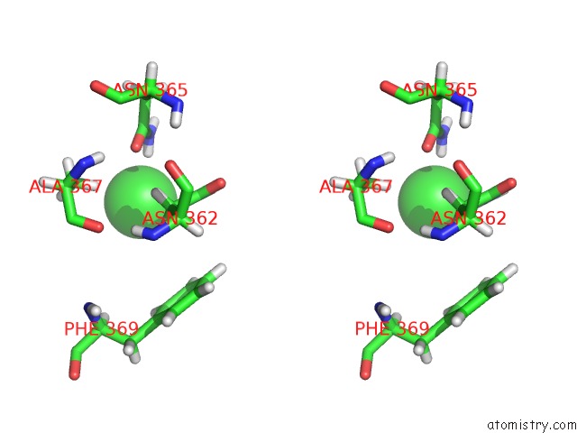
Stereo pair view
Mono view
Stereo pair view
A full contact list of Chlorine with other atoms in the Cl binding
site number 1 of Crystal Structure of Rhdvb P Domain in Complex with Lewis Y within 5.0Å range:
|
Chlorine binding site 2 out of 3 in 4x1x
Go back to
Chlorine binding site 2 out
of 3 in the Crystal Structure of Rhdvb P Domain in Complex with Lewis Y
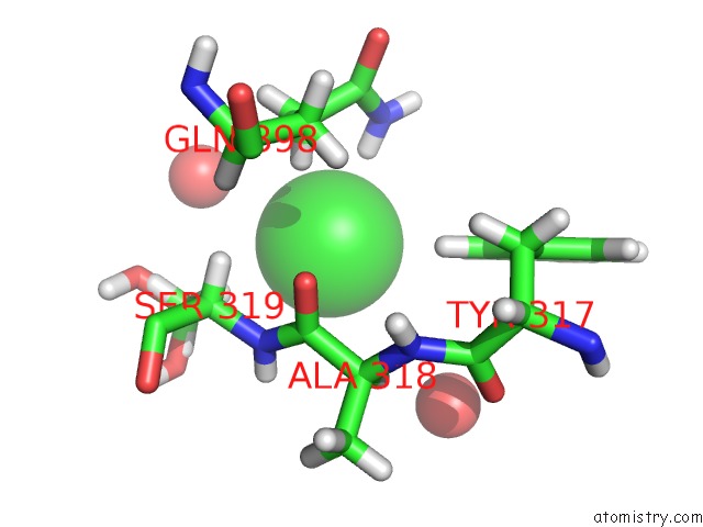
Mono view
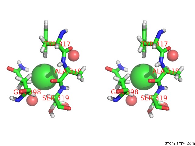
Stereo pair view
Mono view
Stereo pair view
A full contact list of Chlorine with other atoms in the Cl binding
site number 2 of Crystal Structure of Rhdvb P Domain in Complex with Lewis Y within 5.0Å range:
|
Chlorine binding site 3 out of 3 in 4x1x
Go back to
Chlorine binding site 3 out
of 3 in the Crystal Structure of Rhdvb P Domain in Complex with Lewis Y
Mono view
Stereo pair view
Mono view
Stereo pair view
A full contact list of Chlorine with other atoms in the Cl binding
site number 3 of Crystal Structure of Rhdvb P Domain in Complex with Lewis Y within 5.0Å range:
|
Reference:
M.M.Leuthold,
K.P.Dalton,
G.S.Hansman.
Structural Analysis of A Rabbit Hemorrhagic Disease Virus Binding to Histo-Blood Group Antigens. J.Virol. V. 89 2378 2015.
ISSN: ESSN 1098-5514
PubMed: 25505081
DOI: 10.1128/JVI.02832-14
Page generated: Fri Jul 11 22:43:48 2025
ISSN: ESSN 1098-5514
PubMed: 25505081
DOI: 10.1128/JVI.02832-14
Last articles
K in 2WM2K in 2WLN
K in 2WLK
K in 2WLM
K in 2WLO
K in 2WLL
K in 2WLH
K in 2WLI
K in 2WJ6
K in 2WLJ