Chlorine »
PDB 4wxp-4x97 »
4x5p »
Chlorine in PDB 4x5p: Crystal Structure of Fimh in Complex with A Benzoyl-Amidophenyl Alpha- D-Mannopyranoside
Protein crystallography data
The structure of Crystal Structure of Fimh in Complex with A Benzoyl-Amidophenyl Alpha- D-Mannopyranoside, PDB code: 4x5p
was solved by
R.C.Preston,
R.P.Jakob,
B.Fiege,
P.Zihlmann,
S.Rabbani,
O.Schwardt,
X.Jiang,
B.Ernst,
T.Maier,
with X-Ray Crystallography technique. A brief refinement statistics is given in the table below:
| Resolution Low / High (Å) | 38.13 / 1.00 |
| Space group | P 21 21 21 |
| Cell size a, b, c (Å), α, β, γ (°) | 48.840, 55.890, 61.000, 90.00, 90.00, 90.00 |
| R / Rfree (%) | 12.3 / 13.9 |
Chlorine Binding Sites:
The binding sites of Chlorine atom in the Crystal Structure of Fimh in Complex with A Benzoyl-Amidophenyl Alpha- D-Mannopyranoside
(pdb code 4x5p). This binding sites where shown within
5.0 Angstroms radius around Chlorine atom.
In total only one binding site of Chlorine was determined in the Crystal Structure of Fimh in Complex with A Benzoyl-Amidophenyl Alpha- D-Mannopyranoside, PDB code: 4x5p:
In total only one binding site of Chlorine was determined in the Crystal Structure of Fimh in Complex with A Benzoyl-Amidophenyl Alpha- D-Mannopyranoside, PDB code: 4x5p:
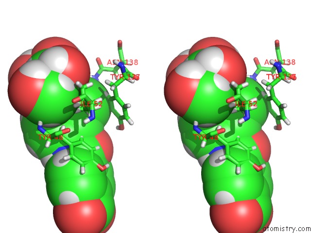
Chlorine binding site 1 out of 1 in 4x5p
Go back to
Chlorine binding site 1 out
of 1 in the Crystal Structure of Fimh in Complex with A Benzoyl-Amidophenyl Alpha- D-Mannopyranoside
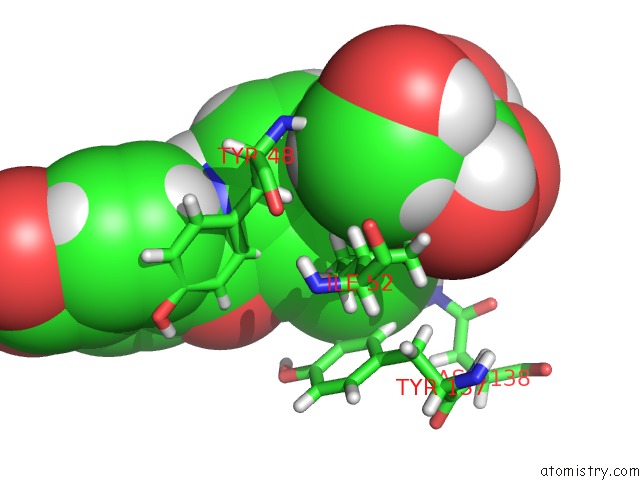
Mono view
Stereo pair view
Mono view
Stereo pair view
A full contact list of Chlorine with other atoms in the Cl binding
site number 1 of Crystal Structure of Fimh in Complex with A Benzoyl-Amidophenyl Alpha- D-Mannopyranoside within 5.0Å range:
|
Reference:
B.Fiege,
S.Rabbani,
R.C.Preston,
R.P.Jakob,
P.Zihlmann,
O.Schwardt,
X.Jiang,
T.Maier,
B.Ernst.
The Tyrosine Gate of the Bacterial Lectin Fimh: A Conformational Analysis By uc(Nmr) Spectroscopy and X-Ray Crystallography. Chembiochem V. 16 1235 2015.
ISSN: ESSN 1439-7633
PubMed: 25940742
DOI: 10.1002/CBIC.201402714
Page generated: Fri Jul 26 03:13:41 2024
ISSN: ESSN 1439-7633
PubMed: 25940742
DOI: 10.1002/CBIC.201402714
Last articles
Zn in 9MJ5Zn in 9HNW
Zn in 9G0L
Zn in 9FNE
Zn in 9DZN
Zn in 9E0I
Zn in 9D32
Zn in 9DAK
Zn in 8ZXC
Zn in 8ZUF