Chlorine »
PDB 4wxp-4x97 »
4x90 »
Chlorine in PDB 4x90: Crystal Structure of Lysosomal Phospholipase A2
Protein crystallography data
The structure of Crystal Structure of Lysosomal Phospholipase A2, PDB code: 4x90
was solved by
A.Glukhova,
J.J.G.Tesmer,
with X-Ray Crystallography technique. A brief refinement statistics is given in the table below:
| Resolution Low / High (Å) | 30.00 / 1.84 |
| Space group | P 1 |
| Cell size a, b, c (Å), α, β, γ (°) | 62.806, 91.151, 100.266, 78.13, 88.46, 88.50 |
| R / Rfree (%) | 15.4 / 17.3 |
Chlorine Binding Sites:
The binding sites of Chlorine atom in the Crystal Structure of Lysosomal Phospholipase A2
(pdb code 4x90). This binding sites where shown within
5.0 Angstroms radius around Chlorine atom.
In total 4 binding sites of Chlorine where determined in the Crystal Structure of Lysosomal Phospholipase A2, PDB code: 4x90:
Jump to Chlorine binding site number: 1; 2; 3; 4;
In total 4 binding sites of Chlorine where determined in the Crystal Structure of Lysosomal Phospholipase A2, PDB code: 4x90:
Jump to Chlorine binding site number: 1; 2; 3; 4;
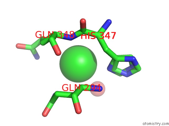
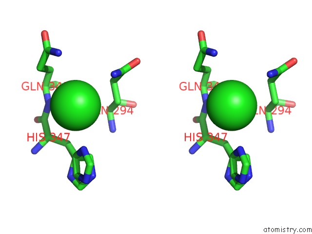
Chlorine binding site 1 out of 4 in 4x90
Go back to
Chlorine binding site 1 out
of 4 in the Crystal Structure of Lysosomal Phospholipase A2
Mono view

Stereo pair view
Mono view
Stereo pair view
A full contact list of Chlorine with other atoms in the Cl binding
site number 1 of Crystal Structure of Lysosomal Phospholipase A2 within 5.0Å range:
|
Chlorine binding site 2 out of 4 in 4x90
Go back to
Chlorine binding site 2 out
of 4 in the Crystal Structure of Lysosomal Phospholipase A2

Mono view

Stereo pair view
Mono view
Stereo pair view
A full contact list of Chlorine with other atoms in the Cl binding
site number 2 of Crystal Structure of Lysosomal Phospholipase A2 within 5.0Å range:
|
Chlorine binding site 3 out of 4 in 4x90
Go back to
Chlorine binding site 3 out
of 4 in the Crystal Structure of Lysosomal Phospholipase A2

Mono view
Stereo pair view
Mono view
Stereo pair view
A full contact list of Chlorine with other atoms in the Cl binding
site number 3 of Crystal Structure of Lysosomal Phospholipase A2 within 5.0Å range:
|
Chlorine binding site 4 out of 4 in 4x90
Go back to
Chlorine binding site 4 out
of 4 in the Crystal Structure of Lysosomal Phospholipase A2

Mono view

Stereo pair view
Mono view
Stereo pair view
A full contact list of Chlorine with other atoms in the Cl binding
site number 4 of Crystal Structure of Lysosomal Phospholipase A2 within 5.0Å range:
|
Reference:
A.Glukhova,
V.Hinkovska-Galcheva,
R.Kelly,
A.Abe,
J.A.Shayman,
J.J.G.Tesmer.
Structure and Function of Lysosomal Phospholipase A2 and Lecithin:Cholesterol Acyltransferase Nat Commun 2015.
ISSN: ESSN 2041-1723
DOI: 10.1038/NCOMMS7250
Page generated: Fri Jul 11 22:49:10 2025
ISSN: ESSN 2041-1723
DOI: 10.1038/NCOMMS7250
Last articles
Mg in 2A6EMg in 2A5Z
Mg in 2A5L
Mg in 2A5Y
Mg in 2A5J
Mg in 2A43
Mg in 2A5G
Mg in 2A5D
Mg in 2A5F
Mg in 2A42