Chlorine »
PDB 4xpf-4y14 »
4xyp »
Chlorine in PDB 4xyp: Crystal Structure of A Piscine Viral Fusion Protein
Protein crystallography data
The structure of Crystal Structure of A Piscine Viral Fusion Protein, PDB code: 4xyp
was solved by
J.D.Cook,
J.E.Lee,
with X-Ray Crystallography technique. A brief refinement statistics is given in the table below:
| Resolution Low / High (Å) | 38.71 / 2.10 |
| Space group | H 3 2 |
| Cell size a, b, c (Å), α, β, γ (°) | 42.874, 42.874, 232.245, 90.00, 90.00, 120.00 |
| R / Rfree (%) | 19.6 / 25.1 |
Chlorine Binding Sites:
The binding sites of Chlorine atom in the Crystal Structure of A Piscine Viral Fusion Protein
(pdb code 4xyp). This binding sites where shown within
5.0 Angstroms radius around Chlorine atom.
In total 2 binding sites of Chlorine where determined in the Crystal Structure of A Piscine Viral Fusion Protein, PDB code: 4xyp:
Jump to Chlorine binding site number: 1; 2;
In total 2 binding sites of Chlorine where determined in the Crystal Structure of A Piscine Viral Fusion Protein, PDB code: 4xyp:
Jump to Chlorine binding site number: 1; 2;
Chlorine binding site 1 out of 2 in 4xyp
Go back to
Chlorine binding site 1 out
of 2 in the Crystal Structure of A Piscine Viral Fusion Protein
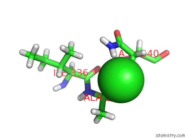
Mono view
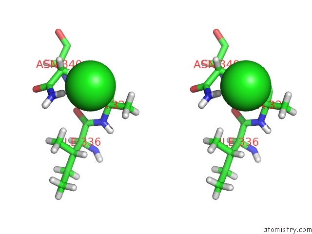
Stereo pair view
Mono view
Stereo pair view
A full contact list of Chlorine with other atoms in the Cl binding
site number 1 of Crystal Structure of A Piscine Viral Fusion Protein within 5.0Å range:
|
Chlorine binding site 2 out of 2 in 4xyp
Go back to
Chlorine binding site 2 out
of 2 in the Crystal Structure of A Piscine Viral Fusion Protein
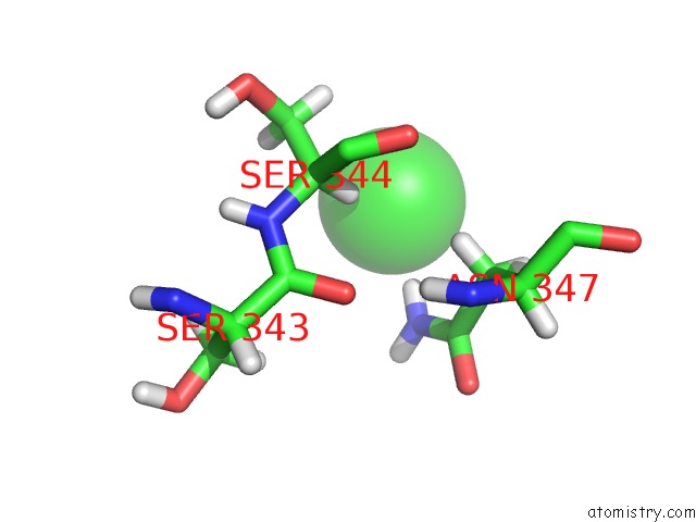
Mono view

Stereo pair view
Mono view
Stereo pair view
A full contact list of Chlorine with other atoms in the Cl binding
site number 2 of Crystal Structure of A Piscine Viral Fusion Protein within 5.0Å range:
|
Reference:
J.D.Cook,
H.Soto-Montoya,
M.K.Korpela,
J.E.Lee.
Electrostatic Architecture of the Infectious Salmon Anemia Virus (Isav) Core Fusion Protein Illustrates A Carboxyl-Carboxylate pH Sensor. J.Biol.Chem. V. 290 18495 2015.
ISSN: ESSN 1083-351X
PubMed: 26082488
DOI: 10.1074/JBC.M115.644781
Page generated: Fri Jul 11 23:07:43 2025
ISSN: ESSN 1083-351X
PubMed: 26082488
DOI: 10.1074/JBC.M115.644781
Last articles
Mg in 3L8ZMg in 3L8Y
Mg in 3L8G
Mg in 3L8F
Mg in 3L7Y
Mg in 3L86
Mg in 3L4P
Mg in 3L6T
Mg in 3L6Q
Mg in 3L2U