Chlorine »
PDB 4yli-4ywy »
4ysh »
Chlorine in PDB 4ysh: Crystal Structure of Glycine Oxidase From Geobacillus Kaustophilus
Enzymatic activity of Crystal Structure of Glycine Oxidase From Geobacillus Kaustophilus
All present enzymatic activity of Crystal Structure of Glycine Oxidase From Geobacillus Kaustophilus:
1.4.3.19;
1.4.3.19;
Protein crystallography data
The structure of Crystal Structure of Glycine Oxidase From Geobacillus Kaustophilus, PDB code: 4ysh
was solved by
T.Shiono,
T.Nomura,
R.Arai,
with X-Ray Crystallography technique. A brief refinement statistics is given in the table below:
| Resolution Low / High (Å) | 50.00 / 2.20 |
| Space group | P 65 2 2 |
| Cell size a, b, c (Å), α, β, γ (°) | 87.945, 87.945, 413.456, 90.00, 90.00, 120.00 |
| R / Rfree (%) | 23.3 / 26.6 |
Chlorine Binding Sites:
The binding sites of Chlorine atom in the Crystal Structure of Glycine Oxidase From Geobacillus Kaustophilus
(pdb code 4ysh). This binding sites where shown within
5.0 Angstroms radius around Chlorine atom.
In total only one binding site of Chlorine was determined in the Crystal Structure of Glycine Oxidase From Geobacillus Kaustophilus, PDB code: 4ysh:
In total only one binding site of Chlorine was determined in the Crystal Structure of Glycine Oxidase From Geobacillus Kaustophilus, PDB code: 4ysh:
Chlorine binding site 1 out of 1 in 4ysh
Go back to
Chlorine binding site 1 out
of 1 in the Crystal Structure of Glycine Oxidase From Geobacillus Kaustophilus
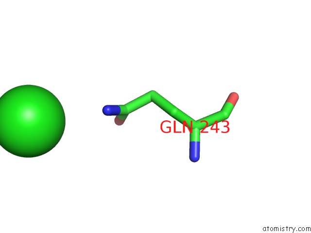
Mono view
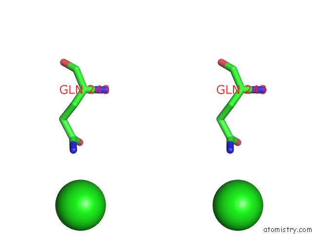
Stereo pair view
Mono view
Stereo pair view
A full contact list of Chlorine with other atoms in the Cl binding
site number 1 of Crystal Structure of Glycine Oxidase From Geobacillus Kaustophilus within 5.0Å range:
|
Reference:
T.Shiono,
R.Arai,
Y.Nishiya,
T.Nomura.
Crystal Structure of Glycine Oxidase From Geobacillus Kaustophilus To Be Published.
Page generated: Fri Jul 11 23:29:24 2025
Last articles
Fe in 9ILTFe in 9IMN
Fe in 9IL4
Fe in 9IKI
Fe in 9IKH
Fe in 9IAB
Fe in 9IKG
Fe in 9IIG
Fe in 9IAC
Fe in 9IKF