Chlorine »
PDB 4z6v-4zg7 »
4zba »
Chlorine in PDB 4zba: Crystal Structure of the Glutathione Transferase URE2P8 From Phanerochaete Chrysosporium with Oxidized Glutathione.
Protein crystallography data
The structure of Crystal Structure of the Glutathione Transferase URE2P8 From Phanerochaete Chrysosporium with Oxidized Glutathione., PDB code: 4zba
was solved by
T.Roret,
C.Didierjean,
with X-Ray Crystallography technique. A brief refinement statistics is given in the table below:
| Resolution Low / High (Å) | 46.30 / 1.50 |
| Space group | P 21 21 21 |
| Cell size a, b, c (Å), α, β, γ (°) | 53.641, 91.872, 180.014, 90.00, 90.00, 90.00 |
| R / Rfree (%) | 15 / 16.9 |
Chlorine Binding Sites:
The binding sites of Chlorine atom in the Crystal Structure of the Glutathione Transferase URE2P8 From Phanerochaete Chrysosporium with Oxidized Glutathione.
(pdb code 4zba). This binding sites where shown within
5.0 Angstroms radius around Chlorine atom.
In total 5 binding sites of Chlorine where determined in the Crystal Structure of the Glutathione Transferase URE2P8 From Phanerochaete Chrysosporium with Oxidized Glutathione., PDB code: 4zba:
Jump to Chlorine binding site number: 1; 2; 3; 4; 5;
In total 5 binding sites of Chlorine where determined in the Crystal Structure of the Glutathione Transferase URE2P8 From Phanerochaete Chrysosporium with Oxidized Glutathione., PDB code: 4zba:
Jump to Chlorine binding site number: 1; 2; 3; 4; 5;
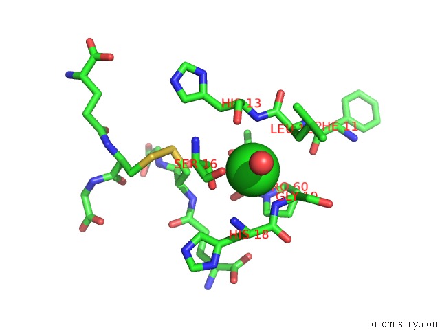
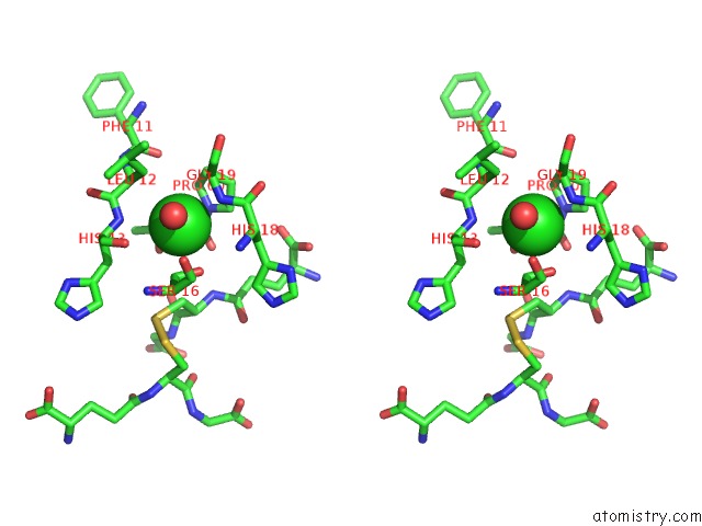
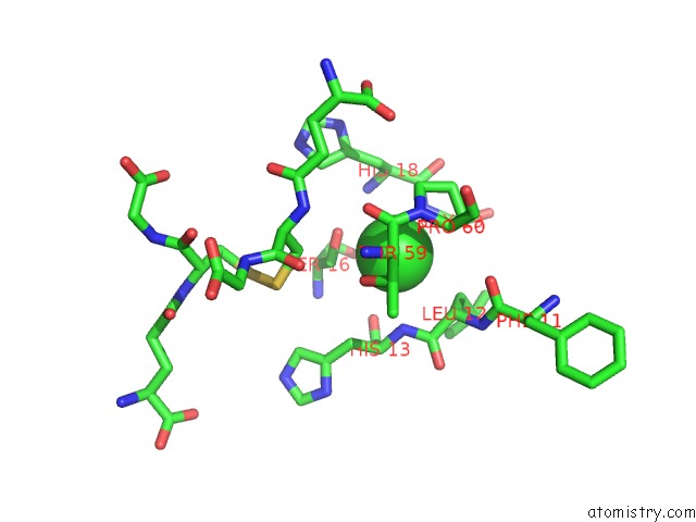
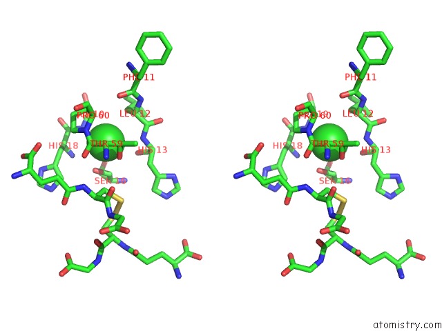
Chlorine binding site 1 out of 5 in 4zba
Go back to
Chlorine binding site 1 out
of 5 in the Crystal Structure of the Glutathione Transferase URE2P8 From Phanerochaete Chrysosporium with Oxidized Glutathione.
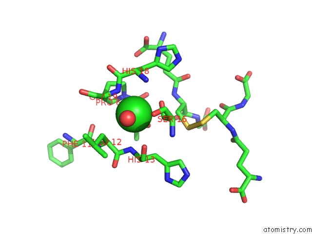
Mono view
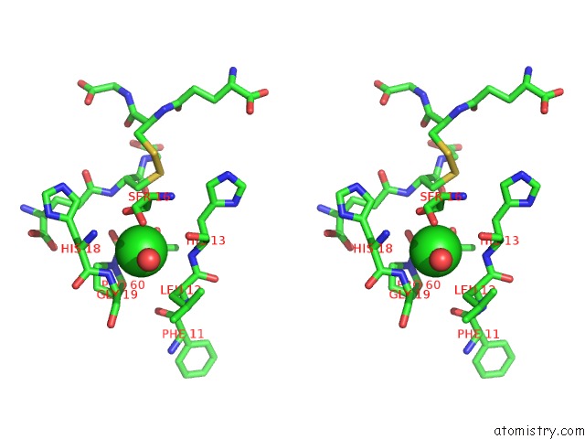
Stereo pair view
Mono view
Stereo pair view
A full contact list of Chlorine with other atoms in the Cl binding
site number 1 of Crystal Structure of the Glutathione Transferase URE2P8 From Phanerochaete Chrysosporium with Oxidized Glutathione. within 5.0Å range:
|
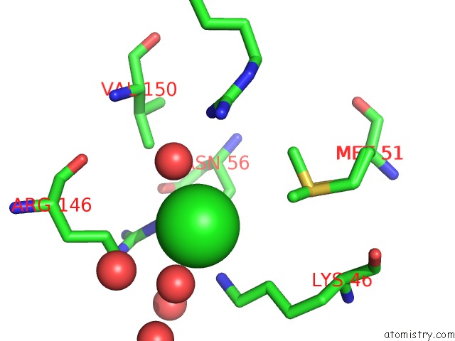
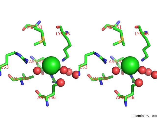
Chlorine binding site 2 out of 5 in 4zba
Go back to
Chlorine binding site 2 out
of 5 in the Crystal Structure of the Glutathione Transferase URE2P8 From Phanerochaete Chrysosporium with Oxidized Glutathione.
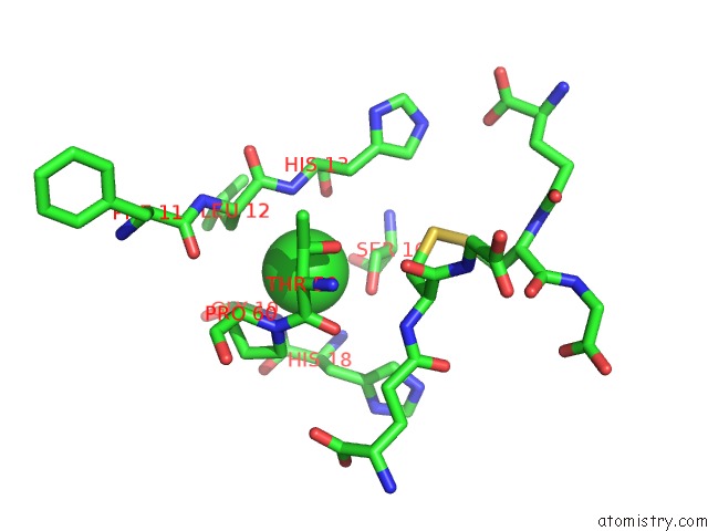
Mono view
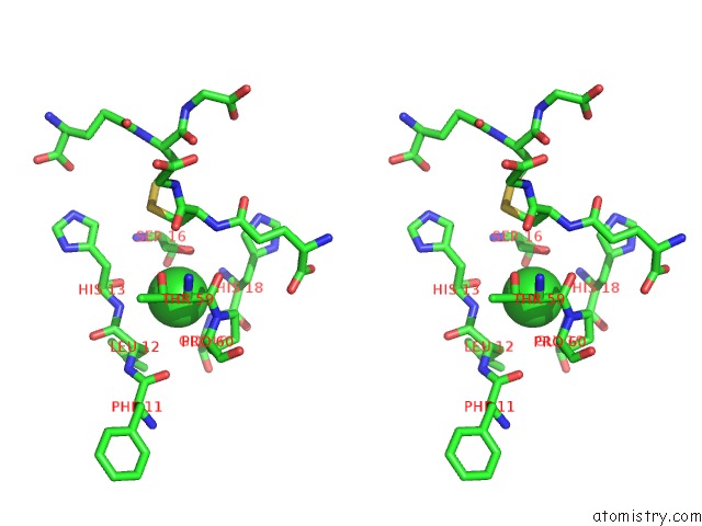
Stereo pair view
Mono view
Stereo pair view
A full contact list of Chlorine with other atoms in the Cl binding
site number 2 of Crystal Structure of the Glutathione Transferase URE2P8 From Phanerochaete Chrysosporium with Oxidized Glutathione. within 5.0Å range:
|
Chlorine binding site 3 out of 5 in 4zba
Go back to
Chlorine binding site 3 out
of 5 in the Crystal Structure of the Glutathione Transferase URE2P8 From Phanerochaete Chrysosporium with Oxidized Glutathione.
Mono view
Stereo pair view
Mono view
Stereo pair view
A full contact list of Chlorine with other atoms in the Cl binding
site number 3 of Crystal Structure of the Glutathione Transferase URE2P8 From Phanerochaete Chrysosporium with Oxidized Glutathione. within 5.0Å range:
|
Chlorine binding site 4 out of 5 in 4zba
Go back to
Chlorine binding site 4 out
of 5 in the Crystal Structure of the Glutathione Transferase URE2P8 From Phanerochaete Chrysosporium with Oxidized Glutathione.
Mono view
Stereo pair view
Mono view
Stereo pair view
A full contact list of Chlorine with other atoms in the Cl binding
site number 4 of Crystal Structure of the Glutathione Transferase URE2P8 From Phanerochaete Chrysosporium with Oxidized Glutathione. within 5.0Å range:
|
Chlorine binding site 5 out of 5 in 4zba
Go back to
Chlorine binding site 5 out
of 5 in the Crystal Structure of the Glutathione Transferase URE2P8 From Phanerochaete Chrysosporium with Oxidized Glutathione.
Mono view
Stereo pair view
Mono view
Stereo pair view
A full contact list of Chlorine with other atoms in the Cl binding
site number 5 of Crystal Structure of the Glutathione Transferase URE2P8 From Phanerochaete Chrysosporium with Oxidized Glutathione. within 5.0Å range:
|
Reference:
T.Roret,
A.Thuillier,
F.Favier,
E.Gelhaye,
C.Didierjean,
M.Morel-Rouhier.
Evolutionary Divergence of URE2PA Glutathione Transferases in Wood Degrading Fungi. Fungal Genet. Biol. V. 83 103 2015.
ISSN: ISSN 1096-0937
PubMed: 26348000
DOI: 10.1016/J.FGB.2015.09.002
Page generated: Fri Jul 11 23:40:07 2025
ISSN: ISSN 1096-0937
PubMed: 26348000
DOI: 10.1016/J.FGB.2015.09.002
Last articles
Mg in 6POPMg in 6PJW
Mg in 6PMO
Mg in 6PMJ
Mg in 6PMI
Mg in 6PLC
Mg in 6PL8
Mg in 6PL7
Mg in 6PKZ
Mg in 6PJV