Chlorine »
PDB 5boz-5bvf »
5bt3 »
Chlorine in PDB 5bt3: Crystal Structure of EP300 Bromodomain in Complex with Sgc-CBP30 Chemical Probe
Enzymatic activity of Crystal Structure of EP300 Bromodomain in Complex with Sgc-CBP30 Chemical Probe
All present enzymatic activity of Crystal Structure of EP300 Bromodomain in Complex with Sgc-CBP30 Chemical Probe:
2.3.1.48;
2.3.1.48;
Protein crystallography data
The structure of Crystal Structure of EP300 Bromodomain in Complex with Sgc-CBP30 Chemical Probe, PDB code: 5bt3
was solved by
C.Tallant,
D.Hay,
T.Krojer,
G.Nunez-Alonso,
S.Picaud,
J.A.Newman,
O.Fedorov,
F.Von Delft,
C.H.Arrowsmith,
A.M.Edwards,
C.Bountra,
P.E.Brennan,
S.Knapp,
Structural Genomics Consortium (Sgc),
with X-Ray Crystallography technique. A brief refinement statistics is given in the table below:
| Resolution Low / High (Å) | 46.27 / 1.05 |
| Space group | P 61 |
| Cell size a, b, c (Å), α, β, γ (°) | 53.431, 53.431, 77.003, 90.00, 90.00, 120.00 |
| R / Rfree (%) | 16.5 / 17.8 |
Chlorine Binding Sites:
The binding sites of Chlorine atom in the Crystal Structure of EP300 Bromodomain in Complex with Sgc-CBP30 Chemical Probe
(pdb code 5bt3). This binding sites where shown within
5.0 Angstroms radius around Chlorine atom.
In total only one binding site of Chlorine was determined in the Crystal Structure of EP300 Bromodomain in Complex with Sgc-CBP30 Chemical Probe, PDB code: 5bt3:
In total only one binding site of Chlorine was determined in the Crystal Structure of EP300 Bromodomain in Complex with Sgc-CBP30 Chemical Probe, PDB code: 5bt3:
Chlorine binding site 1 out of 1 in 5bt3
Go back to
Chlorine binding site 1 out
of 1 in the Crystal Structure of EP300 Bromodomain in Complex with Sgc-CBP30 Chemical Probe
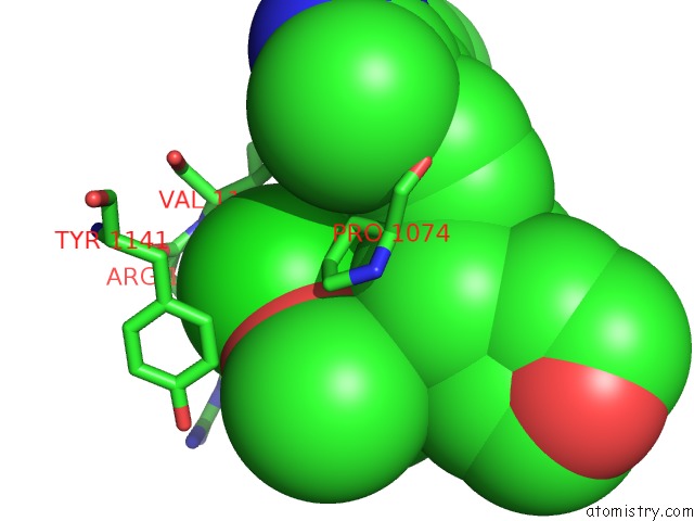
Mono view
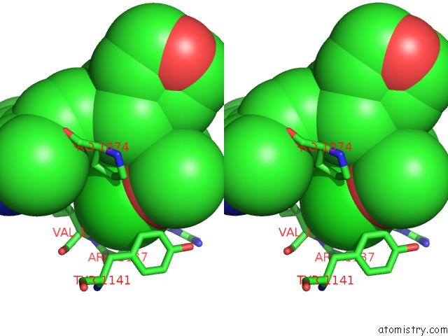
Stereo pair view
Mono view
Stereo pair view
A full contact list of Chlorine with other atoms in the Cl binding
site number 1 of Crystal Structure of EP300 Bromodomain in Complex with Sgc-CBP30 Chemical Probe within 5.0Å range:
|
Reference:
C.Tallant,
D.Hay,
T.Krojer,
G.Nunez-Alonso,
S.Picaud,
J.A.Newman,
O.Fedorov,
F.Von Delft,
C.H.Arrowsmith,
A.M.Edwards,
C.Bountra,
P.E.Brennan,
S.Knapp.
Crystal Structure of EP300 Bromodomain in Complex with A 3,5-Dimethylisoxazol Ligand To Be Published.
Page generated: Sat Jul 12 00:30:14 2025
Last articles
Mg in 5TFGMg in 5TFF
Mg in 5TFJ
Mg in 5TFI
Mg in 5TDS
Mg in 5TFD
Mg in 5TFC
Mg in 5TFB
Mg in 5TFA
Mg in 5TF0