Chlorine »
PDB 5dab-5dij »
5dar »
Chlorine in PDB 5dar: Crystal Structure of the Base of the Ribosomal P Stalk From Methanococcus Jannaschii
Protein crystallography data
The structure of Crystal Structure of the Base of the Ribosomal P Stalk From Methanococcus Jannaschii, PDB code: 5dar
was solved by
A.G.Gabdulkhakov,
I.V.Mitroshin,
M.B.Garber,
with X-Ray Crystallography technique. A brief refinement statistics is given in the table below:
| Resolution Low / High (Å) | 20.00 / 2.90 |
| Space group | P 1 21 1 |
| Cell size a, b, c (Å), α, β, γ (°) | 72.396, 88.451, 95.231, 90.00, 102.19, 90.00 |
| R / Rfree (%) | 26.4 / 29.7 |
Other elements in 5dar:
The structure of Crystal Structure of the Base of the Ribosomal P Stalk From Methanococcus Jannaschii also contains other interesting chemical elements:
| Magnesium | (Mg) | 8 atoms |
| Potassium | (K) | 5 atoms |
Chlorine Binding Sites:
The binding sites of Chlorine atom in the Crystal Structure of the Base of the Ribosomal P Stalk From Methanococcus Jannaschii
(pdb code 5dar). This binding sites where shown within
5.0 Angstroms radius around Chlorine atom.
In total 3 binding sites of Chlorine where determined in the Crystal Structure of the Base of the Ribosomal P Stalk From Methanococcus Jannaschii, PDB code: 5dar:
Jump to Chlorine binding site number: 1; 2; 3;
In total 3 binding sites of Chlorine where determined in the Crystal Structure of the Base of the Ribosomal P Stalk From Methanococcus Jannaschii, PDB code: 5dar:
Jump to Chlorine binding site number: 1; 2; 3;
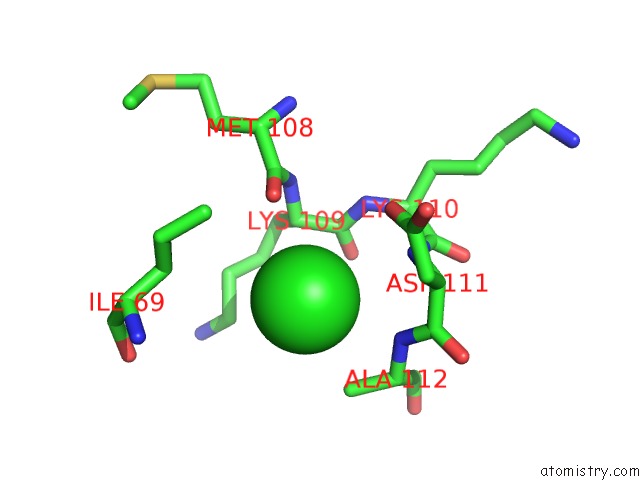
Chlorine binding site 1 out of 3 in 5dar
Go back to
Chlorine binding site 1 out
of 3 in the Crystal Structure of the Base of the Ribosomal P Stalk From Methanococcus Jannaschii
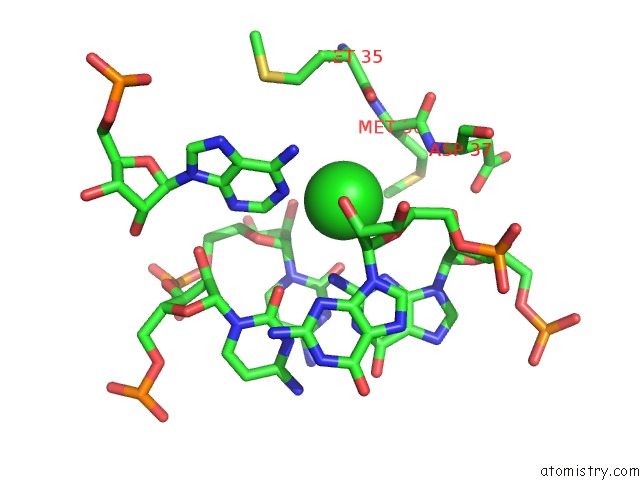
Mono view
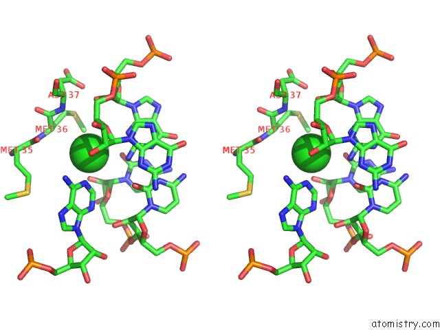
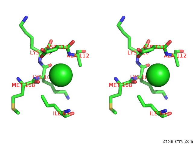
Stereo pair view
Mono view
Stereo pair view
A full contact list of Chlorine with other atoms in the Cl binding
site number 1 of Crystal Structure of the Base of the Ribosomal P Stalk From Methanococcus Jannaschii within 5.0Å range:
|
Chlorine binding site 2 out of 3 in 5dar
Go back to
Chlorine binding site 2 out
of 3 in the Crystal Structure of the Base of the Ribosomal P Stalk From Methanococcus Jannaschii
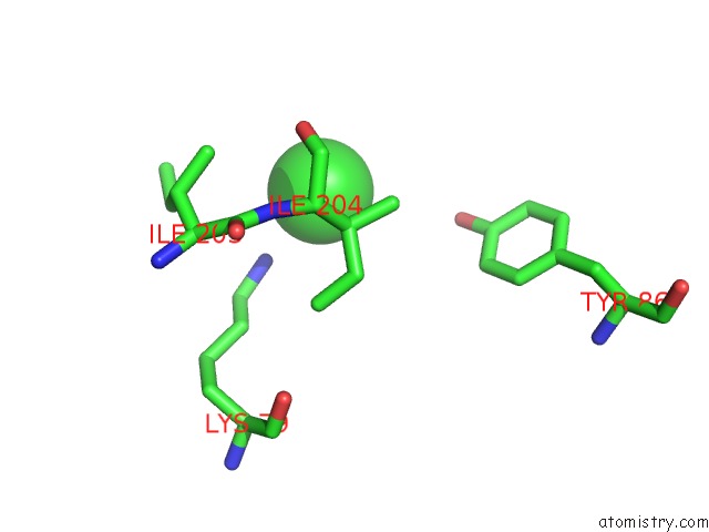
Mono view
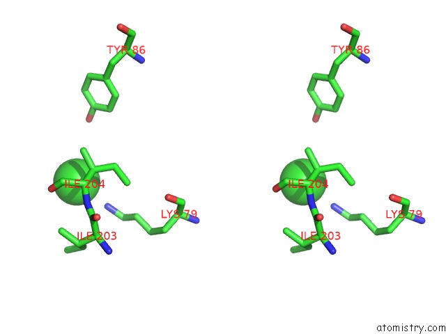
Stereo pair view
Mono view
Stereo pair view
A full contact list of Chlorine with other atoms in the Cl binding
site number 2 of Crystal Structure of the Base of the Ribosomal P Stalk From Methanococcus Jannaschii within 5.0Å range:
|
Chlorine binding site 3 out of 3 in 5dar
Go back to
Chlorine binding site 3 out
of 3 in the Crystal Structure of the Base of the Ribosomal P Stalk From Methanococcus Jannaschii
Mono view
Stereo pair view
Mono view
Stereo pair view
A full contact list of Chlorine with other atoms in the Cl binding
site number 3 of Crystal Structure of the Base of the Ribosomal P Stalk From Methanococcus Jannaschii within 5.0Å range:
|
Reference:
A.G.Gabdulkhakov,
I.V.Mitroshin,
M.B.Garber.
Crystal Structure of the Base of the Ribosomal P Stalk From Methanococcus Jannaschii To Be Published.
Page generated: Sat Jul 12 01:10:58 2025
Last articles
Mg in 5G0RMg in 5G5V
Mg in 5G3T
Mg in 5G5T
Mg in 5G5S
Mg in 5G4A
Mg in 5G57
Mg in 5G50
Mg in 5G41
Mg in 5G3Z