Chlorine »
PDB 5e20-5eae »
5e78 »
Chlorine in PDB 5e78: Crystal Structure of P450 BM3 Heme Domain Variant Complexed with Co(III)Sep
Enzymatic activity of Crystal Structure of P450 BM3 Heme Domain Variant Complexed with Co(III)Sep
All present enzymatic activity of Crystal Structure of P450 BM3 Heme Domain Variant Complexed with Co(III)Sep:
1.14.14.1; 1.6.2.4;
1.14.14.1; 1.6.2.4;
Protein crystallography data
The structure of Crystal Structure of P450 BM3 Heme Domain Variant Complexed with Co(III)Sep, PDB code: 5e78
was solved by
S.Panneerselvm,
A.Shehzad,
M.Bocola,
J.Mueller-Dieckmann,
U.Schwaneberg,
with X-Ray Crystallography technique. A brief refinement statistics is given in the table below:
| Resolution Low / High (Å) | 20.00 / 2.00 |
| Space group | P 21 21 21 |
| Cell size a, b, c (Å), α, β, γ (°) | 58.970, 128.540, 150.060, 90.00, 90.00, 90.00 |
| R / Rfree (%) | 17.5 / 21.2 |
Other elements in 5e78:
The structure of Crystal Structure of P450 BM3 Heme Domain Variant Complexed with Co(III)Sep also contains other interesting chemical elements:
| Cobalt | (Co) | 1 atom |
| Iron | (Fe) | 2 atoms |
Chlorine Binding Sites:
The binding sites of Chlorine atom in the Crystal Structure of P450 BM3 Heme Domain Variant Complexed with Co(III)Sep
(pdb code 5e78). This binding sites where shown within
5.0 Angstroms radius around Chlorine atom.
In total only one binding site of Chlorine was determined in the Crystal Structure of P450 BM3 Heme Domain Variant Complexed with Co(III)Sep, PDB code: 5e78:
In total only one binding site of Chlorine was determined in the Crystal Structure of P450 BM3 Heme Domain Variant Complexed with Co(III)Sep, PDB code: 5e78:
Chlorine binding site 1 out of 1 in 5e78
Go back to
Chlorine binding site 1 out
of 1 in the Crystal Structure of P450 BM3 Heme Domain Variant Complexed with Co(III)Sep
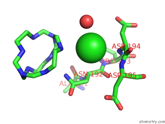
Mono view
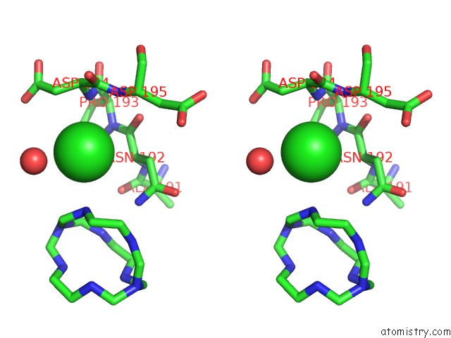
Stereo pair view
Mono view
Stereo pair view
A full contact list of Chlorine with other atoms in the Cl binding
site number 1 of Crystal Structure of P450 BM3 Heme Domain Variant Complexed with Co(III)Sep within 5.0Å range:
|
Reference:
S.Panneerselvam,
A.Shehzad,
J.Mueller-Dieckmann,
M.Wilmanns,
M.Bocola,
M.D.Davari,
U.Schwaneberg.
Crystallographic Insights Into A Cobalt (III) Sepulchrate Based Alternative Cofactor System of P450 BM3 Monooxygenase. Biochim. Biophys. Acta V.1866 134 2018.
ISSN: ISSN 0006-3002
PubMed: 28739446
DOI: 10.1016/J.BBAPAP.2017.07.010
Page generated: Sat Jul 12 01:32:31 2025
ISSN: ISSN 0006-3002
PubMed: 28739446
DOI: 10.1016/J.BBAPAP.2017.07.010
Last articles
K in 5E33K in 5E2Q
K in 5E1A
K in 5DWX
K in 5DUN
K in 5DSX
K in 5DRY
K in 5DRT
K in 5DMJ
K in 5DQK