Chlorine »
PDB 5grn-5h4z »
5h0z »
Chlorine in PDB 5h0z: Crystal Structure of P113A Mutated Human Transthyretin
Protein crystallography data
The structure of Crystal Structure of P113A Mutated Human Transthyretin, PDB code: 5h0z
was solved by
T.Yokoyama,
Y.Hanawa,
T.Obita,
M.Mizuguchi,
with X-Ray Crystallography technique. A brief refinement statistics is given in the table below:
| Resolution Low / High (Å) | 32.13 / 1.74 |
| Space group | I 2 2 2 |
| Cell size a, b, c (Å), α, β, γ (°) | 41.180, 64.260, 85.370, 90.00, 90.00, 90.00 |
| R / Rfree (%) | 22.6 / 25.7 |
Chlorine Binding Sites:
The binding sites of Chlorine atom in the Crystal Structure of P113A Mutated Human Transthyretin
(pdb code 5h0z). This binding sites where shown within
5.0 Angstroms radius around Chlorine atom.
In total only one binding site of Chlorine was determined in the Crystal Structure of P113A Mutated Human Transthyretin, PDB code: 5h0z:
In total only one binding site of Chlorine was determined in the Crystal Structure of P113A Mutated Human Transthyretin, PDB code: 5h0z:
Chlorine binding site 1 out of 1 in 5h0z
Go back to
Chlorine binding site 1 out
of 1 in the Crystal Structure of P113A Mutated Human Transthyretin
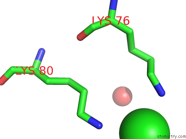
Mono view
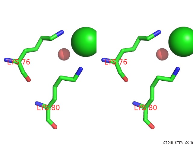
Stereo pair view
Mono view
Stereo pair view
A full contact list of Chlorine with other atoms in the Cl binding
site number 1 of Crystal Structure of P113A Mutated Human Transthyretin within 5.0Å range:
|
Reference:
T.Yokoyama,
Y.Hanawa,
T.Obita,
M.Mizuguchi.
Stability and Crystal Structures of HIS88 Mutant Human Transthyretins Febs Lett. V. 591 1862 2017.
ISSN: ISSN 1873-3468
PubMed: 28563699
DOI: 10.1002/1873-3468.12704
Page generated: Sat Jul 12 02:40:25 2025
ISSN: ISSN 1873-3468
PubMed: 28563699
DOI: 10.1002/1873-3468.12704
Last articles
Na in 1I0BNa in 1HZY
Na in 1HZC
Na in 1HZB
Na in 1HTW
Na in 1HYA
Na in 1HXN
Na in 1HVX
Na in 1HNF
Na in 1HN1