Chlorine »
PDB 5hqa-5i1f »
5hsv »
Chlorine in PDB 5hsv: X-Ray Structure of A Cypa-Alisporivir Complex at 1.5 Angstrom Resolution
Enzymatic activity of X-Ray Structure of A Cypa-Alisporivir Complex at 1.5 Angstrom Resolution
All present enzymatic activity of X-Ray Structure of A Cypa-Alisporivir Complex at 1.5 Angstrom Resolution:
5.2.1.8;
5.2.1.8;
Protein crystallography data
The structure of X-Ray Structure of A Cypa-Alisporivir Complex at 1.5 Angstrom Resolution, PDB code: 5hsv
was solved by
M.Dujardin,
J.Bouckaert,
P.Rucktooa,
X.Hanoulle,
with X-Ray Crystallography technique. A brief refinement statistics is given in the table below:
| Resolution Low / High (Å) | 36.25 / 1.50 |
| Space group | P 1 |
| Cell size a, b, c (Å), α, β, γ (°) | 37.904, 68.443, 71.952, 115.12, 103.17, 93.96 |
| R / Rfree (%) | 16.6 / 19 |
Chlorine Binding Sites:
The binding sites of Chlorine atom in the X-Ray Structure of A Cypa-Alisporivir Complex at 1.5 Angstrom Resolution
(pdb code 5hsv). This binding sites where shown within
5.0 Angstroms radius around Chlorine atom.
In total 4 binding sites of Chlorine where determined in the X-Ray Structure of A Cypa-Alisporivir Complex at 1.5 Angstrom Resolution, PDB code: 5hsv:
Jump to Chlorine binding site number: 1; 2; 3; 4;
In total 4 binding sites of Chlorine where determined in the X-Ray Structure of A Cypa-Alisporivir Complex at 1.5 Angstrom Resolution, PDB code: 5hsv:
Jump to Chlorine binding site number: 1; 2; 3; 4;
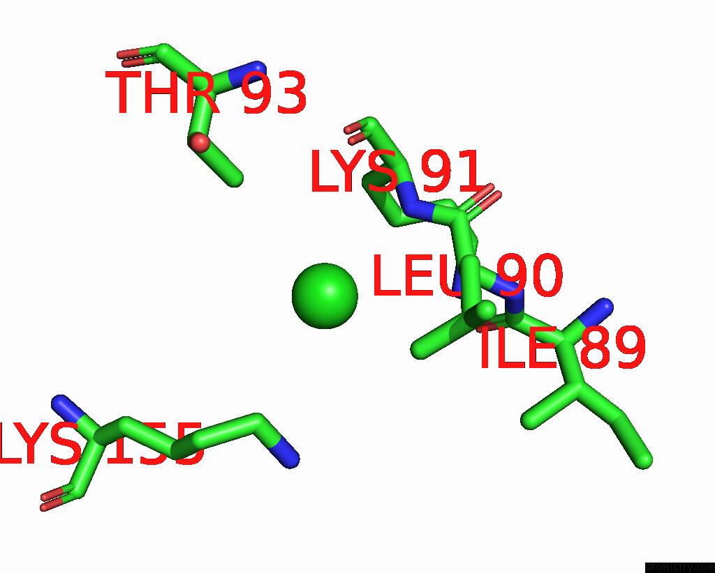
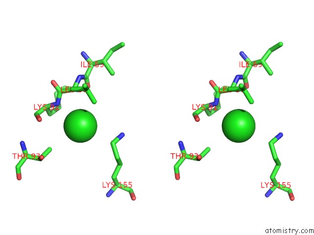
Chlorine binding site 1 out of 4 in 5hsv
Go back to
Chlorine binding site 1 out
of 4 in the X-Ray Structure of A Cypa-Alisporivir Complex at 1.5 Angstrom Resolution
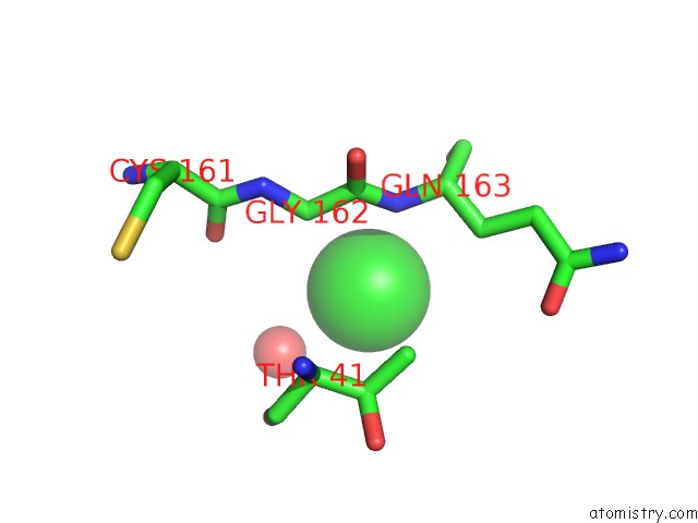
Mono view
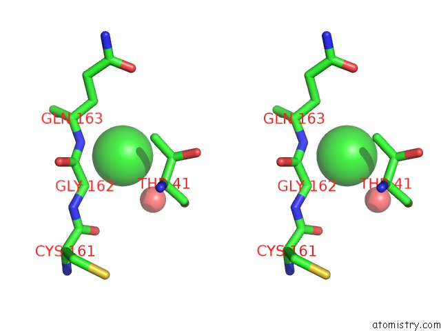
Stereo pair view
Mono view
Stereo pair view
A full contact list of Chlorine with other atoms in the Cl binding
site number 1 of X-Ray Structure of A Cypa-Alisporivir Complex at 1.5 Angstrom Resolution within 5.0Å range:
|
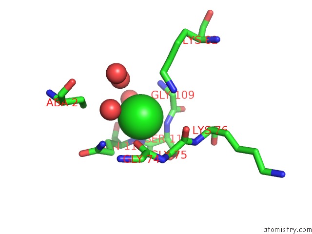
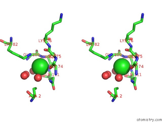
Chlorine binding site 2 out of 4 in 5hsv
Go back to
Chlorine binding site 2 out
of 4 in the X-Ray Structure of A Cypa-Alisporivir Complex at 1.5 Angstrom Resolution
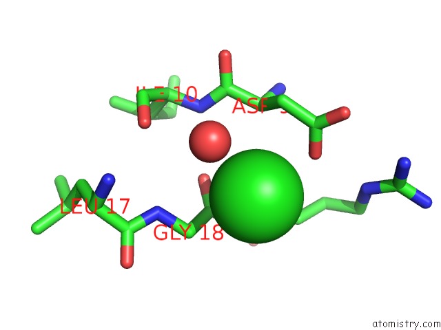
Mono view
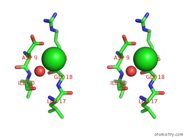
Stereo pair view
Mono view
Stereo pair view
A full contact list of Chlorine with other atoms in the Cl binding
site number 2 of X-Ray Structure of A Cypa-Alisporivir Complex at 1.5 Angstrom Resolution within 5.0Å range:
|
Chlorine binding site 3 out of 4 in 5hsv
Go back to
Chlorine binding site 3 out
of 4 in the X-Ray Structure of A Cypa-Alisporivir Complex at 1.5 Angstrom Resolution
Mono view
Stereo pair view
Mono view
Stereo pair view
A full contact list of Chlorine with other atoms in the Cl binding
site number 3 of X-Ray Structure of A Cypa-Alisporivir Complex at 1.5 Angstrom Resolution within 5.0Å range:
|
Chlorine binding site 4 out of 4 in 5hsv
Go back to
Chlorine binding site 4 out
of 4 in the X-Ray Structure of A Cypa-Alisporivir Complex at 1.5 Angstrom Resolution
Mono view
Stereo pair view
Mono view
Stereo pair view
A full contact list of Chlorine with other atoms in the Cl binding
site number 4 of X-Ray Structure of A Cypa-Alisporivir Complex at 1.5 Angstrom Resolution within 5.0Å range:
|
Reference:
M.Dujardin,
J.Bouckaert,
P.Rucktooa,
X.Hanoulle.
X-Ray Structure of Alisporivir in Complex with Cyclophilin A at 1.5 Angstrom Resolution. Acta Crystallogr F Struct V. 74 583 2018BIOL Commun.
ISSN: ESSN 2053-230X
PubMed: 30198892
DOI: 10.1107/S2053230X18010415
Page generated: Sat Jul 12 02:53:47 2025
ISSN: ESSN 2053-230X
PubMed: 30198892
DOI: 10.1107/S2053230X18010415
Last articles
Mg in 2ACXMg in 2A9R
Mg in 2A9K
Mg in 2A9F
Mg in 2A84
Mg in 2A87
Mg in 2A6H
Mg in 2A6R
Mg in 2A78
Mg in 2A7Q