Chlorine »
PDB 5imx-5ivr »
5ipe »
Chlorine in PDB 5ipe: Human Histidine Triad Nucleotide Binding Protein 1 (HHINT1) Nucleoside Thiophosphoramidate Catalytic Product Complex
Protein crystallography data
The structure of Human Histidine Triad Nucleotide Binding Protein 1 (HHINT1) Nucleoside Thiophosphoramidate Catalytic Product Complex, PDB code: 5ipe
was solved by
K.M.Maize,
B.C.Finzel,
with X-Ray Crystallography technique. A brief refinement statistics is given in the table below:
| Resolution Low / High (Å) | 39.82 / 1.45 |
| Space group | C 1 2 1 |
| Cell size a, b, c (Å), α, β, γ (°) | 78.270, 46.311, 63.963, 90.00, 94.72, 90.00 |
| R / Rfree (%) | 15.9 / 18.1 |
Chlorine Binding Sites:
The binding sites of Chlorine atom in the Human Histidine Triad Nucleotide Binding Protein 1 (HHINT1) Nucleoside Thiophosphoramidate Catalytic Product Complex
(pdb code 5ipe). This binding sites where shown within
5.0 Angstroms radius around Chlorine atom.
In total only one binding site of Chlorine was determined in the Human Histidine Triad Nucleotide Binding Protein 1 (HHINT1) Nucleoside Thiophosphoramidate Catalytic Product Complex, PDB code: 5ipe:
In total only one binding site of Chlorine was determined in the Human Histidine Triad Nucleotide Binding Protein 1 (HHINT1) Nucleoside Thiophosphoramidate Catalytic Product Complex, PDB code: 5ipe:
Chlorine binding site 1 out of 1 in 5ipe
Go back to
Chlorine binding site 1 out
of 1 in the Human Histidine Triad Nucleotide Binding Protein 1 (HHINT1) Nucleoside Thiophosphoramidate Catalytic Product Complex

Mono view
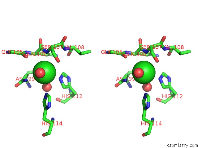
Stereo pair view
Mono view
Stereo pair view
A full contact list of Chlorine with other atoms in the Cl binding
site number 1 of Human Histidine Triad Nucleotide Binding Protein 1 (HHINT1) Nucleoside Thiophosphoramidate Catalytic Product Complex within 5.0Å range:
|
Reference:
R.Shah,
K.M.Maize,
X.Zhou,
B.C.Finzel,
C.R.Wagner.
Caught Before Released: Structural Mapping of the Reaction Trajectory For the Sofosbuvir Activating Enzyme, Human Histidine Triad Nucleotide Binding Protein 1 (HHINT1). Biochemistry V. 56 3559 2017.
ISSN: ISSN 1520-4995
PubMed: 28691797
DOI: 10.1021/ACS.BIOCHEM.7B00148
Page generated: Sat Jul 12 03:14:23 2025
ISSN: ISSN 1520-4995
PubMed: 28691797
DOI: 10.1021/ACS.BIOCHEM.7B00148
Last articles
Mg in 7EWKMg in 7F0L
Mg in 7F7A
Mg in 7F6N
Mg in 7F75
Mg in 7F6J
Mg in 7F6E
Mg in 7F6F
Mg in 7F50
Mg in 7F68