Chlorine »
PDB 5jkd-5jsv »
5jpi »
Chlorine in PDB 5jpi: 2.15 Angstrom Crystal Structure of S-Adenosylhomocysteinase From Cryptosporidium Parvum in Complex with D-Eritadenine and Nad
Enzymatic activity of 2.15 Angstrom Crystal Structure of S-Adenosylhomocysteinase From Cryptosporidium Parvum in Complex with D-Eritadenine and Nad
All present enzymatic activity of 2.15 Angstrom Crystal Structure of S-Adenosylhomocysteinase From Cryptosporidium Parvum in Complex with D-Eritadenine and Nad:
3.3.1.1;
3.3.1.1;
Protein crystallography data
The structure of 2.15 Angstrom Crystal Structure of S-Adenosylhomocysteinase From Cryptosporidium Parvum in Complex with D-Eritadenine and Nad, PDB code: 5jpi
was solved by
G.Minasov,
L.Shuvalova,
O.Kiryukhina,
I.Dubrovska,
B.Bishop,
K.Kwon,
W.F.Anderson,
Center For Structural Genomics Of Infectious Diseases(Csgid),
with X-Ray Crystallography technique. A brief refinement statistics is given in the table below:
| Resolution Low / High (Å) | 29.97 / 2.15 |
| Space group | P 21 21 21 |
| Cell size a, b, c (Å), α, β, γ (°) | 115.847, 121.218, 178.770, 90.00, 90.00, 90.00 |
| R / Rfree (%) | 14.2 / 18.1 |
Chlorine Binding Sites:
The binding sites of Chlorine atom in the 2.15 Angstrom Crystal Structure of S-Adenosylhomocysteinase From Cryptosporidium Parvum in Complex with D-Eritadenine and Nad
(pdb code 5jpi). This binding sites where shown within
5.0 Angstroms radius around Chlorine atom.
In total 4 binding sites of Chlorine where determined in the 2.15 Angstrom Crystal Structure of S-Adenosylhomocysteinase From Cryptosporidium Parvum in Complex with D-Eritadenine and Nad, PDB code: 5jpi:
Jump to Chlorine binding site number: 1; 2; 3; 4;
In total 4 binding sites of Chlorine where determined in the 2.15 Angstrom Crystal Structure of S-Adenosylhomocysteinase From Cryptosporidium Parvum in Complex with D-Eritadenine and Nad, PDB code: 5jpi:
Jump to Chlorine binding site number: 1; 2; 3; 4;
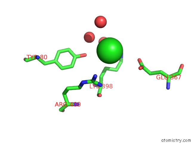
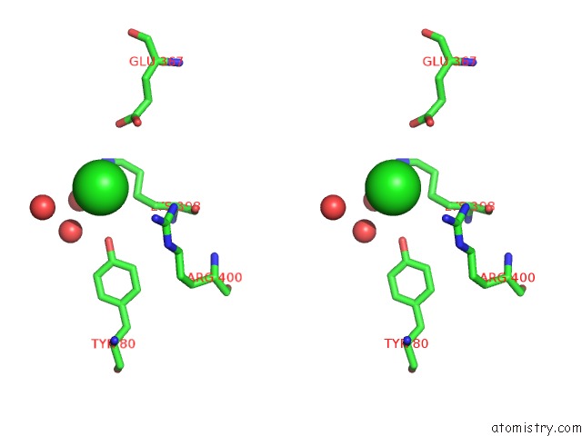
Chlorine binding site 1 out of 4 in 5jpi
Go back to
Chlorine binding site 1 out
of 4 in the 2.15 Angstrom Crystal Structure of S-Adenosylhomocysteinase From Cryptosporidium Parvum in Complex with D-Eritadenine and Nad
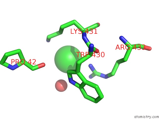
Mono view
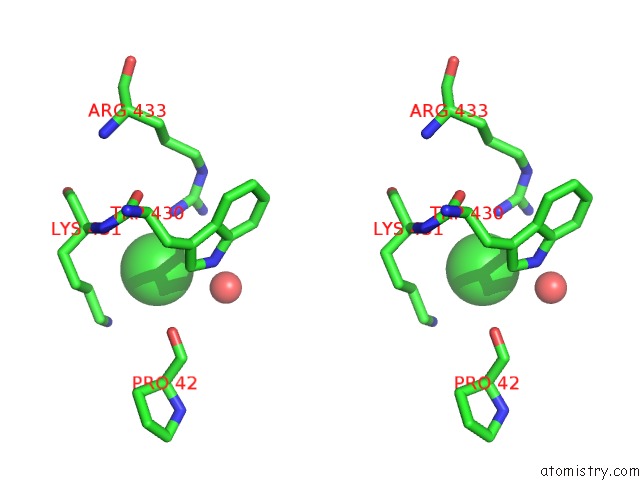
Stereo pair view
Mono view
Stereo pair view
A full contact list of Chlorine with other atoms in the Cl binding
site number 1 of 2.15 Angstrom Crystal Structure of S-Adenosylhomocysteinase From Cryptosporidium Parvum in Complex with D-Eritadenine and Nad within 5.0Å range:
|
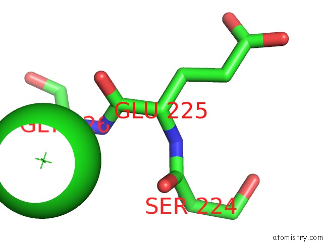
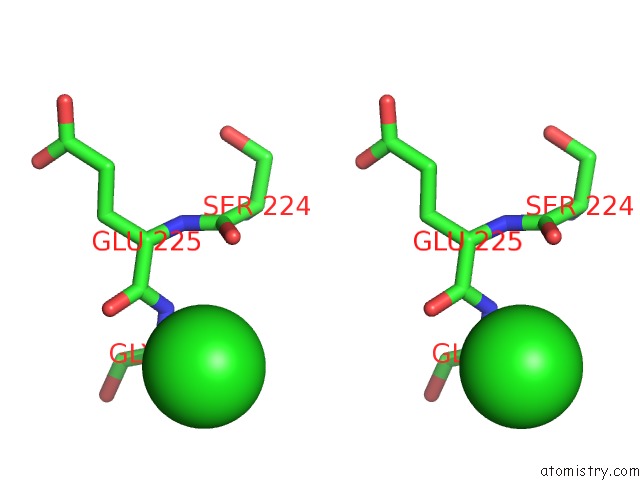
Chlorine binding site 2 out of 4 in 5jpi
Go back to
Chlorine binding site 2 out
of 4 in the 2.15 Angstrom Crystal Structure of S-Adenosylhomocysteinase From Cryptosporidium Parvum in Complex with D-Eritadenine and Nad
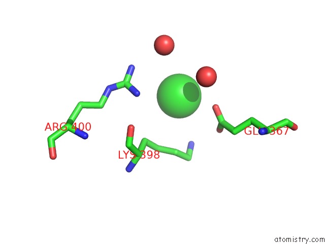
Mono view
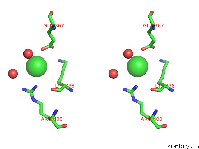
Stereo pair view
Mono view
Stereo pair view
A full contact list of Chlorine with other atoms in the Cl binding
site number 2 of 2.15 Angstrom Crystal Structure of S-Adenosylhomocysteinase From Cryptosporidium Parvum in Complex with D-Eritadenine and Nad within 5.0Å range:
|
Chlorine binding site 3 out of 4 in 5jpi
Go back to
Chlorine binding site 3 out
of 4 in the 2.15 Angstrom Crystal Structure of S-Adenosylhomocysteinase From Cryptosporidium Parvum in Complex with D-Eritadenine and Nad
Mono view
Stereo pair view
Mono view
Stereo pair view
A full contact list of Chlorine with other atoms in the Cl binding
site number 3 of 2.15 Angstrom Crystal Structure of S-Adenosylhomocysteinase From Cryptosporidium Parvum in Complex with D-Eritadenine and Nad within 5.0Å range:
|
Chlorine binding site 4 out of 4 in 5jpi
Go back to
Chlorine binding site 4 out
of 4 in the 2.15 Angstrom Crystal Structure of S-Adenosylhomocysteinase From Cryptosporidium Parvum in Complex with D-Eritadenine and Nad
Mono view
Stereo pair view
Mono view
Stereo pair view
A full contact list of Chlorine with other atoms in the Cl binding
site number 4 of 2.15 Angstrom Crystal Structure of S-Adenosylhomocysteinase From Cryptosporidium Parvum in Complex with D-Eritadenine and Nad within 5.0Å range:
|
Reference:
G.Minasov,
L.Shuvalova,
O.Kiryukhina,
I.Dubrovska,
B.Bishop,
K.Kwon,
W.F.Anderson,
Center For Structural Genomics Of Infectious Diseases(Csgid).
2.15 Angstrom Crystal Structure of S-Adenosylhomocysteinase From Cryptosporidium Parvum in Complex with D-Eritadenine and Nad. To Be Published.
Page generated: Sat Jul 12 03:42:42 2025
Last articles
I in 6AXTI in 6AXV
I in 6AXS
I in 6AN0
I in 6AXR
I in 5ZBA
I in 6A4Y
I in 5ZRF
I in 6A9B
I in 6A9A