Chlorine »
PDB 5kgg-5kp3 »
5kgh »
Chlorine in PDB 5kgh: X-Ray Structure of A Glucosamine N-Acetyltransferase From Clostridium Acetobutylicum, Mutant Y297F
Protein crystallography data
The structure of X-Ray Structure of A Glucosamine N-Acetyltransferase From Clostridium Acetobutylicum, Mutant Y297F, PDB code: 5kgh
was solved by
B.J.Dopkins,
J.B.Thoden,
P.A.Tipton,
H.M.Holden,
with X-Ray Crystallography technique. A brief refinement statistics is given in the table below:
| Resolution Low / High (Å) | 50.00 / 1.80 |
| Space group | P 1 21 1 |
| Cell size a, b, c (Å), α, β, γ (°) | 64.294, 65.941, 90.449, 90.00, 106.57, 90.00 |
| R / Rfree (%) | 20.5 / 24.9 |
Chlorine Binding Sites:
The binding sites of Chlorine atom in the X-Ray Structure of A Glucosamine N-Acetyltransferase From Clostridium Acetobutylicum, Mutant Y297F
(pdb code 5kgh). This binding sites where shown within
5.0 Angstroms radius around Chlorine atom.
In total only one binding site of Chlorine was determined in the X-Ray Structure of A Glucosamine N-Acetyltransferase From Clostridium Acetobutylicum, Mutant Y297F, PDB code: 5kgh:
In total only one binding site of Chlorine was determined in the X-Ray Structure of A Glucosamine N-Acetyltransferase From Clostridium Acetobutylicum, Mutant Y297F, PDB code: 5kgh:
Chlorine binding site 1 out of 1 in 5kgh
Go back to
Chlorine binding site 1 out
of 1 in the X-Ray Structure of A Glucosamine N-Acetyltransferase From Clostridium Acetobutylicum, Mutant Y297F
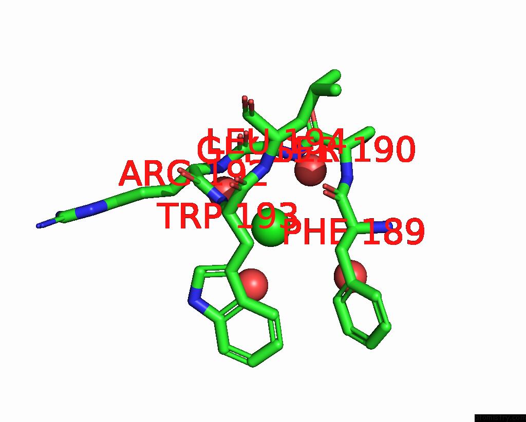
Mono view
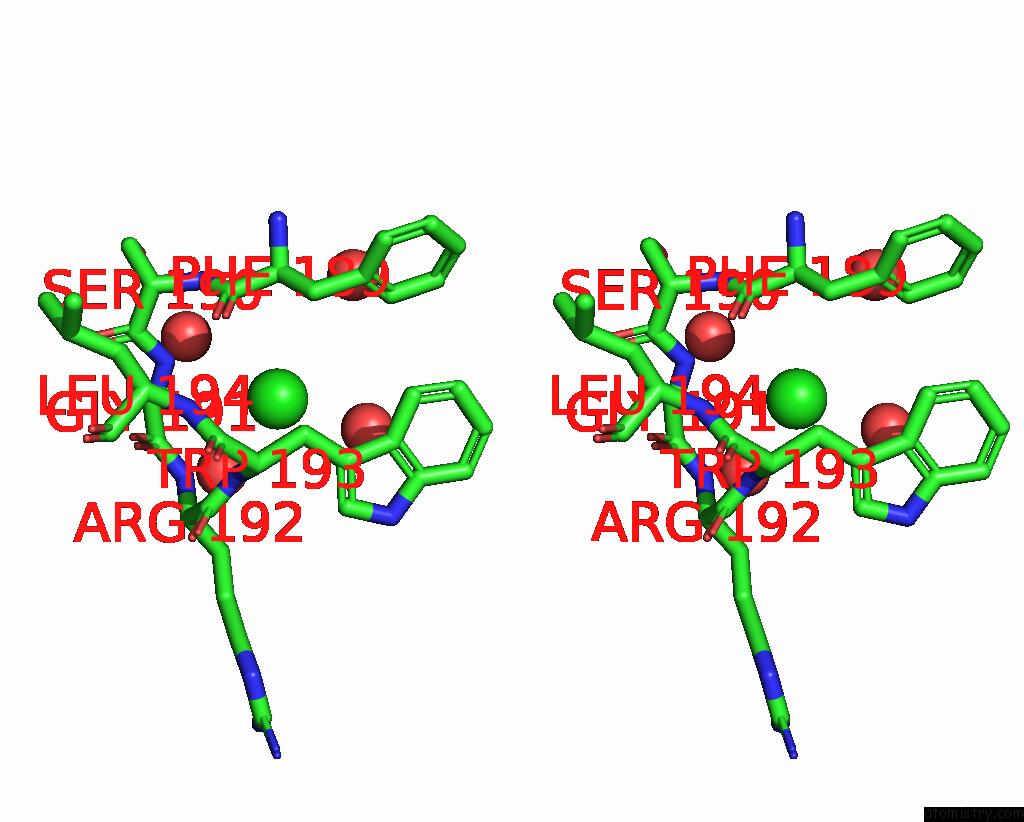
Stereo pair view
Mono view
Stereo pair view
A full contact list of Chlorine with other atoms in the Cl binding
site number 1 of X-Ray Structure of A Glucosamine N-Acetyltransferase From Clostridium Acetobutylicum, Mutant Y297F within 5.0Å range:
|
Reference:
B.J.Dopkins,
P.A.Tipton,
J.B.Thoden,
H.M.Holden.
Structural Studies on A Glucosamine/Glucosaminide N-Acetyltransferase. Biochemistry V. 55 4495 2016.
ISSN: ISSN 0006-2960
PubMed: 27348258
DOI: 10.1021/ACS.BIOCHEM.6B00536
Page generated: Sat Jul 12 04:03:50 2025
ISSN: ISSN 0006-2960
PubMed: 27348258
DOI: 10.1021/ACS.BIOCHEM.6B00536
Last articles
Mg in 7DR0Mg in 7DR1
Mg in 7DU2
Mg in 7DSP
Mg in 7DSJ
Mg in 7DSI
Mg in 7DRP
Mg in 7DSH
Mg in 7DSA
Mg in 7DRX