Chlorine »
PDB 5luj-5lzm »
5luk »
Chlorine in PDB 5luk: Structure of A Double Variant of Cutinase 2 From Thermobifida Cellulosilytica
Protein crystallography data
The structure of Structure of A Double Variant of Cutinase 2 From Thermobifida Cellulosilytica, PDB code: 5luk
was solved by
A.Hromic,
A.Lyskowski,
K.Gruber,
with X-Ray Crystallography technique. A brief refinement statistics is given in the table below:
| Resolution Low / High (Å) | 37.89 / 1.45 |
| Space group | P 21 21 21 |
| Cell size a, b, c (Å), α, β, γ (°) | 40.595, 50.512, 105.477, 90.00, 90.00, 90.00 |
| R / Rfree (%) | 14.4 / 16.4 |
Other elements in 5luk:
The structure of Structure of A Double Variant of Cutinase 2 From Thermobifida Cellulosilytica also contains other interesting chemical elements:
| Magnesium | (Mg) | 2 atoms |
Chlorine Binding Sites:
The binding sites of Chlorine atom in the Structure of A Double Variant of Cutinase 2 From Thermobifida Cellulosilytica
(pdb code 5luk). This binding sites where shown within
5.0 Angstroms radius around Chlorine atom.
In total 3 binding sites of Chlorine where determined in the Structure of A Double Variant of Cutinase 2 From Thermobifida Cellulosilytica, PDB code: 5luk:
Jump to Chlorine binding site number: 1; 2; 3;
In total 3 binding sites of Chlorine where determined in the Structure of A Double Variant of Cutinase 2 From Thermobifida Cellulosilytica, PDB code: 5luk:
Jump to Chlorine binding site number: 1; 2; 3;
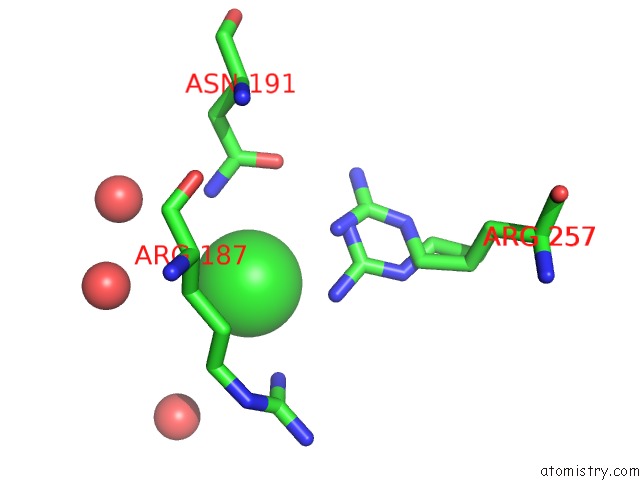
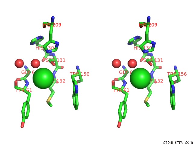
Chlorine binding site 1 out of 3 in 5luk
Go back to
Chlorine binding site 1 out
of 3 in the Structure of A Double Variant of Cutinase 2 From Thermobifida Cellulosilytica
Mono view
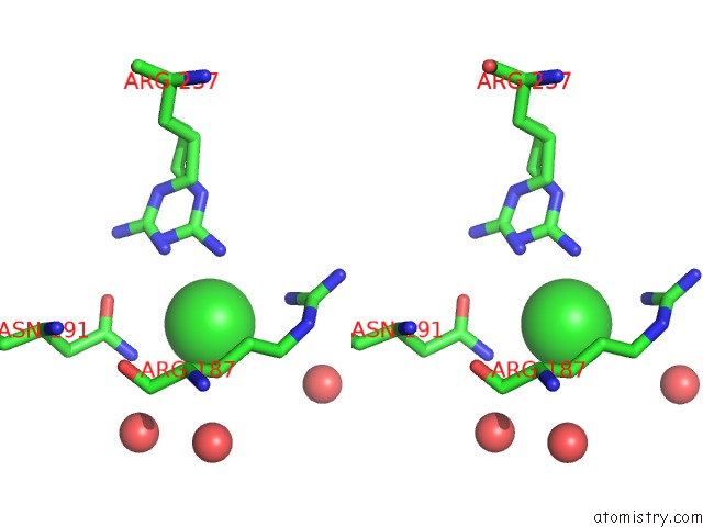
Stereo pair view
Mono view
Stereo pair view
A full contact list of Chlorine with other atoms in the Cl binding
site number 1 of Structure of A Double Variant of Cutinase 2 From Thermobifida Cellulosilytica within 5.0Å range:
|
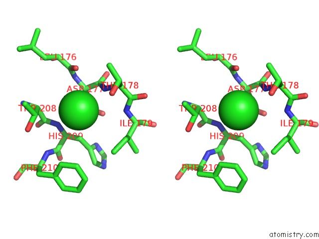
Chlorine binding site 2 out of 3 in 5luk
Go back to
Chlorine binding site 2 out
of 3 in the Structure of A Double Variant of Cutinase 2 From Thermobifida Cellulosilytica

Mono view
Stereo pair view
Mono view
Stereo pair view
A full contact list of Chlorine with other atoms in the Cl binding
site number 2 of Structure of A Double Variant of Cutinase 2 From Thermobifida Cellulosilytica within 5.0Å range:
|
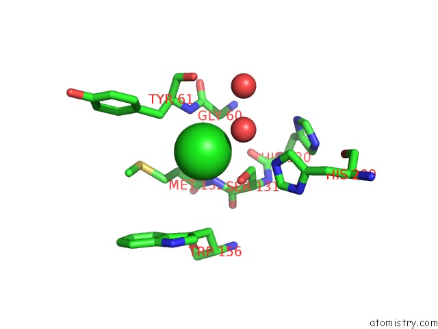
Chlorine binding site 3 out of 3 in 5luk
Go back to
Chlorine binding site 3 out
of 3 in the Structure of A Double Variant of Cutinase 2 From Thermobifida Cellulosilytica
Mono view
Stereo pair view
Mono view
Stereo pair view
A full contact list of Chlorine with other atoms in the Cl binding
site number 3 of Structure of A Double Variant of Cutinase 2 From Thermobifida Cellulosilytica within 5.0Å range:
|
Reference:
D.Ribitsch,
A.Hromic,
S.Zitzenbacher,
B.Zartl,
C.Gamerith,
A.Pellis,
A.Jungbauer,
A.Yskowski,
G.Steinkellner,
K.Gruber,
R.Tscheliessnig,
E.Herrero Acero,
G.M.Guebitz.
Small Cause, Large Effect: Structural Characterization of Cutinases From Thermobifida Cellulosilytica. Biotechnol. Bioeng. V. 114 2481 2017.
ISSN: ESSN 1097-0290
PubMed: 28671263
DOI: 10.1002/BIT.26372
Page generated: Sat Jul 12 05:08:51 2025
ISSN: ESSN 1097-0290
PubMed: 28671263
DOI: 10.1002/BIT.26372
Last articles
Mg in 1R3CMg in 1R2R
Mg in 1R2Q
Mg in 1R2C
Mg in 1R10
Mg in 1R0Z
Mg in 1R0X
Mg in 1R0A
Mg in 1R03
Mg in 1QZR