Chlorine »
PDB 5mlx-5mtr »
5mrs »
Chlorine in PDB 5mrs: Crystal Structure of L1 Protease Lysobacter Sp. XL1 in Complex with Aebsf
Protein crystallography data
The structure of Crystal Structure of L1 Protease Lysobacter Sp. XL1 in Complex with Aebsf, PDB code: 5mrs
was solved by
A.Gabdulkhakov,
S.Tishchenko,
A.Lisov,
A.Leontievsky,
with X-Ray Crystallography technique. A brief refinement statistics is given in the table below:
| Resolution Low / High (Å) | 48.21 / 1.90 |
| Space group | P 1 21 1 |
| Cell size a, b, c (Å), α, β, γ (°) | 41.855, 122.553, 78.988, 90.00, 98.64, 90.00 |
| R / Rfree (%) | 20.1 / 24.2 |
Other elements in 5mrs:
The structure of Crystal Structure of L1 Protease Lysobacter Sp. XL1 in Complex with Aebsf also contains other interesting chemical elements:
| Fluorine | (F) | 3 atoms |
Chlorine Binding Sites:
The binding sites of Chlorine atom in the Crystal Structure of L1 Protease Lysobacter Sp. XL1 in Complex with Aebsf
(pdb code 5mrs). This binding sites where shown within
5.0 Angstroms radius around Chlorine atom.
In total 2 binding sites of Chlorine where determined in the Crystal Structure of L1 Protease Lysobacter Sp. XL1 in Complex with Aebsf, PDB code: 5mrs:
Jump to Chlorine binding site number: 1; 2;
In total 2 binding sites of Chlorine where determined in the Crystal Structure of L1 Protease Lysobacter Sp. XL1 in Complex with Aebsf, PDB code: 5mrs:
Jump to Chlorine binding site number: 1; 2;
Chlorine binding site 1 out of 2 in 5mrs
Go back to
Chlorine binding site 1 out
of 2 in the Crystal Structure of L1 Protease Lysobacter Sp. XL1 in Complex with Aebsf
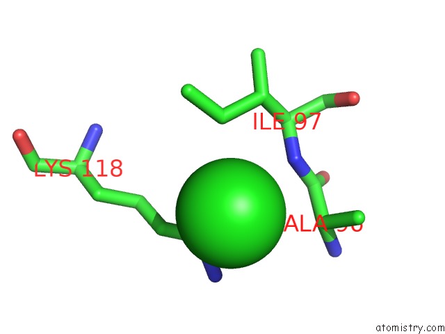
Mono view
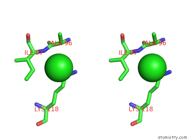
Stereo pair view
Mono view
Stereo pair view
A full contact list of Chlorine with other atoms in the Cl binding
site number 1 of Crystal Structure of L1 Protease Lysobacter Sp. XL1 in Complex with Aebsf within 5.0Å range:
|
Chlorine binding site 2 out of 2 in 5mrs
Go back to
Chlorine binding site 2 out
of 2 in the Crystal Structure of L1 Protease Lysobacter Sp. XL1 in Complex with Aebsf
Mono view
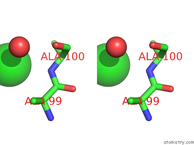
Stereo pair view
Mono view
Stereo pair view
A full contact list of Chlorine with other atoms in the Cl binding
site number 2 of Crystal Structure of L1 Protease Lysobacter Sp. XL1 in Complex with Aebsf within 5.0Å range:
|
Reference:
A.Gabdulkhakov,
S.Tishchenko,
A.Lisov,
A.Leontievsky.
Crystal Structure of L1 Protease Lysobacter Sp. XL1 in Complex with Aebsf To Be Published.
Page generated: Sat Jul 12 05:37:20 2025
Last articles
Mg in 2ZDYMg in 2ZEJ
Mg in 2ZDG
Mg in 2ZCQ
Mg in 2ZCE
Mg in 2ZCR
Mg in 2ZBG
Mg in 2ZCF
Mg in 2Z75
Mg in 2ZBF