Chlorine »
PDB 5mtt-5mzc »
5mvc »
Chlorine in PDB 5mvc: Crystal Structure of Potent Human Dihydroorotate Dehydrogenase Inhibitors Based on Hydroxylated Azole Scaffolds
Enzymatic activity of Crystal Structure of Potent Human Dihydroorotate Dehydrogenase Inhibitors Based on Hydroxylated Azole Scaffolds
All present enzymatic activity of Crystal Structure of Potent Human Dihydroorotate Dehydrogenase Inhibitors Based on Hydroxylated Azole Scaffolds:
1.3.5.2;
1.3.5.2;
Protein crystallography data
The structure of Crystal Structure of Potent Human Dihydroorotate Dehydrogenase Inhibitors Based on Hydroxylated Azole Scaffolds, PDB code: 5mvc
was solved by
P.Goyal,
M.Andersson,
A.C.Moritzer,
S.Sainas,
A.C.Pippione,
D.Boschi,
S.Al-Kadaraghi,
M.Lolli,
R.Friemann,
with X-Ray Crystallography technique. A brief refinement statistics is given in the table below:
| Resolution Low / High (Å) | 29.85 / 1.85 |
| Space group | P 32 2 1 |
| Cell size a, b, c (Å), α, β, γ (°) | 91.190, 91.190, 122.645, 90.00, 90.00, 120.00 |
| R / Rfree (%) | 16.2 / 18.9 |
Other elements in 5mvc:
The structure of Crystal Structure of Potent Human Dihydroorotate Dehydrogenase Inhibitors Based on Hydroxylated Azole Scaffolds also contains other interesting chemical elements:
| Fluorine | (F) | 4 atoms |
Chlorine Binding Sites:
The binding sites of Chlorine atom in the Crystal Structure of Potent Human Dihydroorotate Dehydrogenase Inhibitors Based on Hydroxylated Azole Scaffolds
(pdb code 5mvc). This binding sites where shown within
5.0 Angstroms radius around Chlorine atom.
In total 5 binding sites of Chlorine where determined in the Crystal Structure of Potent Human Dihydroorotate Dehydrogenase Inhibitors Based on Hydroxylated Azole Scaffolds, PDB code: 5mvc:
Jump to Chlorine binding site number: 1; 2; 3; 4; 5;
In total 5 binding sites of Chlorine where determined in the Crystal Structure of Potent Human Dihydroorotate Dehydrogenase Inhibitors Based on Hydroxylated Azole Scaffolds, PDB code: 5mvc:
Jump to Chlorine binding site number: 1; 2; 3; 4; 5;
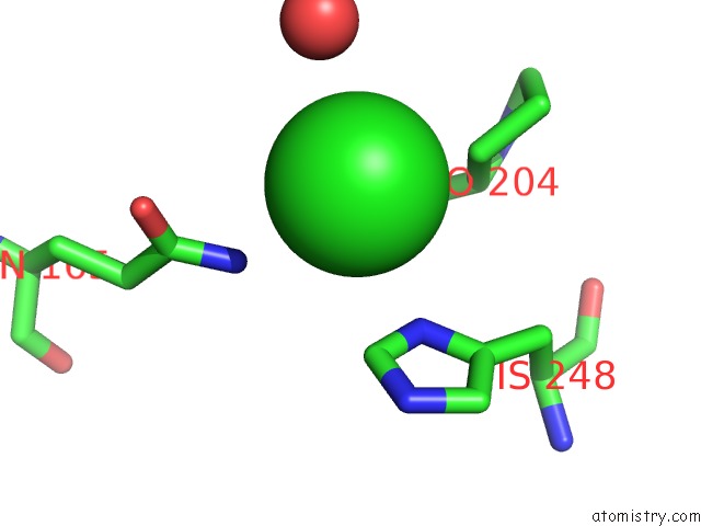
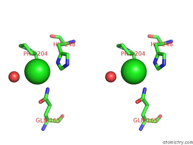
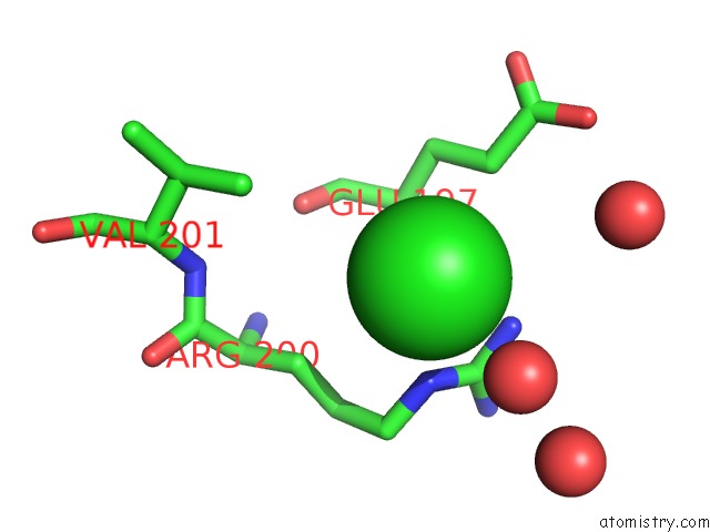
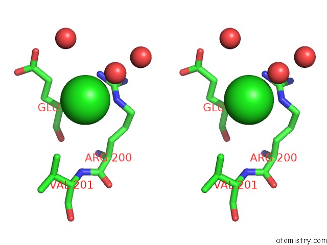
Chlorine binding site 1 out of 5 in 5mvc
Go back to
Chlorine binding site 1 out
of 5 in the Crystal Structure of Potent Human Dihydroorotate Dehydrogenase Inhibitors Based on Hydroxylated Azole Scaffolds
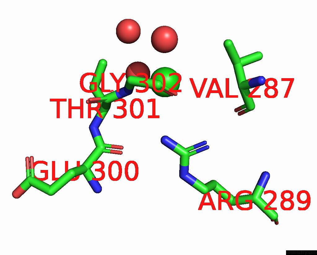
Mono view
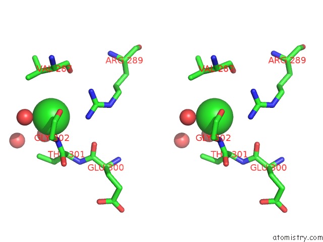
Stereo pair view
Mono view
Stereo pair view
A full contact list of Chlorine with other atoms in the Cl binding
site number 1 of Crystal Structure of Potent Human Dihydroorotate Dehydrogenase Inhibitors Based on Hydroxylated Azole Scaffolds within 5.0Å range:
|
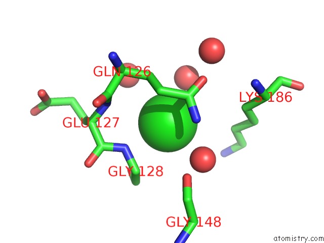
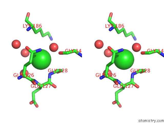
Chlorine binding site 2 out of 5 in 5mvc
Go back to
Chlorine binding site 2 out
of 5 in the Crystal Structure of Potent Human Dihydroorotate Dehydrogenase Inhibitors Based on Hydroxylated Azole Scaffolds
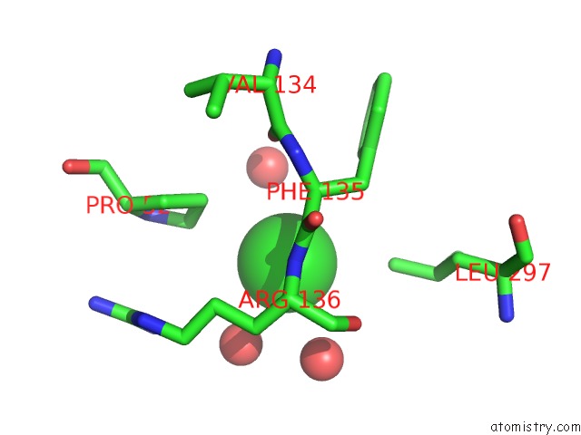
Mono view
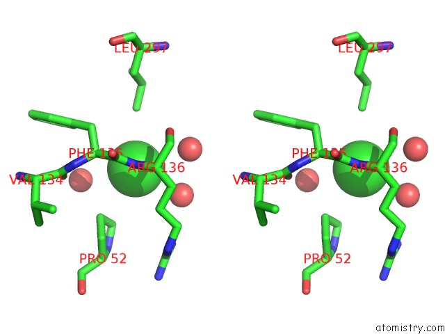
Stereo pair view
Mono view
Stereo pair view
A full contact list of Chlorine with other atoms in the Cl binding
site number 2 of Crystal Structure of Potent Human Dihydroorotate Dehydrogenase Inhibitors Based on Hydroxylated Azole Scaffolds within 5.0Å range:
|
Chlorine binding site 3 out of 5 in 5mvc
Go back to
Chlorine binding site 3 out
of 5 in the Crystal Structure of Potent Human Dihydroorotate Dehydrogenase Inhibitors Based on Hydroxylated Azole Scaffolds
Mono view
Stereo pair view
Mono view
Stereo pair view
A full contact list of Chlorine with other atoms in the Cl binding
site number 3 of Crystal Structure of Potent Human Dihydroorotate Dehydrogenase Inhibitors Based on Hydroxylated Azole Scaffolds within 5.0Å range:
|
Chlorine binding site 4 out of 5 in 5mvc
Go back to
Chlorine binding site 4 out
of 5 in the Crystal Structure of Potent Human Dihydroorotate Dehydrogenase Inhibitors Based on Hydroxylated Azole Scaffolds
Mono view
Stereo pair view
Mono view
Stereo pair view
A full contact list of Chlorine with other atoms in the Cl binding
site number 4 of Crystal Structure of Potent Human Dihydroorotate Dehydrogenase Inhibitors Based on Hydroxylated Azole Scaffolds within 5.0Å range:
|
Chlorine binding site 5 out of 5 in 5mvc
Go back to
Chlorine binding site 5 out
of 5 in the Crystal Structure of Potent Human Dihydroorotate Dehydrogenase Inhibitors Based on Hydroxylated Azole Scaffolds
Mono view
Stereo pair view
Mono view
Stereo pair view
A full contact list of Chlorine with other atoms in the Cl binding
site number 5 of Crystal Structure of Potent Human Dihydroorotate Dehydrogenase Inhibitors Based on Hydroxylated Azole Scaffolds within 5.0Å range:
|
Reference:
S.Sainas,
A.C.Pippione,
M.Giorgis,
E.Lupino,
P.Goyal,
C.Ramondetti,
B.Buccinna,
M.Piccinini,
R.C.Braga,
C.H.Andrade,
M.Andersson,
A.C.Moritzer,
R.Friemann,
S.Mensa,
S.Al-Kadaraghi,
D.Boschi,
M.L.Lolli.
Design, Synthesis, Biological Evaluation and X-Ray Structural Studies of Potent Human Dihydroorotate Dehydrogenase Inhibitors Based on Hydroxylated Azole Scaffolds. Eur J Med Chem V. 129 287 2017.
ISSN: ISSN 1768-3254
PubMed: 28235702
DOI: 10.1016/J.EJMECH.2017.02.017
Page generated: Sat Jul 12 05:40:03 2025
ISSN: ISSN 1768-3254
PubMed: 28235702
DOI: 10.1016/J.EJMECH.2017.02.017
Last articles
Mg in 4LPNMg in 4LNF
Mg in 4LPL
Mg in 4LOC
Mg in 4LOX
Mg in 4LNN
Mg in 4LNU
Mg in 4LNO
Mg in 4LNK
Mg in 4LNL