Chlorine »
PDB 5n75-5ncw »
5na9 »
Chlorine in PDB 5na9: The X-Ray Structure of Bovine Pancreatic Ribonuclease Incubated in the Presence of An Excess of Carboplatin (1:10 Ratio)
Enzymatic activity of The X-Ray Structure of Bovine Pancreatic Ribonuclease Incubated in the Presence of An Excess of Carboplatin (1:10 Ratio)
All present enzymatic activity of The X-Ray Structure of Bovine Pancreatic Ribonuclease Incubated in the Presence of An Excess of Carboplatin (1:10 Ratio):
3.1.27.5;
3.1.27.5;
Protein crystallography data
The structure of The X-Ray Structure of Bovine Pancreatic Ribonuclease Incubated in the Presence of An Excess of Carboplatin (1:10 Ratio), PDB code: 5na9
was solved by
G.Ferraro,
A.Merlino,
with X-Ray Crystallography technique. A brief refinement statistics is given in the table below:
| Resolution Low / High (Å) | 55.57 / 2.07 |
| Space group | P 32 2 1 |
| Cell size a, b, c (Å), α, β, γ (°) | 64.168, 64.168, 64.271, 90.00, 90.00, 120.00 |
| R / Rfree (%) | 17.8 / 24.4 |
Other elements in 5na9:
The structure of The X-Ray Structure of Bovine Pancreatic Ribonuclease Incubated in the Presence of An Excess of Carboplatin (1:10 Ratio) also contains other interesting chemical elements:
| Caesium | (Cs) | 5 atoms |
| Platinum | (Pt) | 2 atoms |
Chlorine Binding Sites:
The binding sites of Chlorine atom in the The X-Ray Structure of Bovine Pancreatic Ribonuclease Incubated in the Presence of An Excess of Carboplatin (1:10 Ratio)
(pdb code 5na9). This binding sites where shown within
5.0 Angstroms radius around Chlorine atom.
In total 9 binding sites of Chlorine where determined in the The X-Ray Structure of Bovine Pancreatic Ribonuclease Incubated in the Presence of An Excess of Carboplatin (1:10 Ratio), PDB code: 5na9:
Jump to Chlorine binding site number: 1; 2; 3; 4; 5; 6; 7; 8; 9;
In total 9 binding sites of Chlorine where determined in the The X-Ray Structure of Bovine Pancreatic Ribonuclease Incubated in the Presence of An Excess of Carboplatin (1:10 Ratio), PDB code: 5na9:
Jump to Chlorine binding site number: 1; 2; 3; 4; 5; 6; 7; 8; 9;
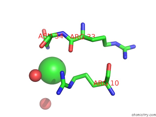
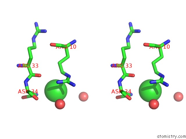
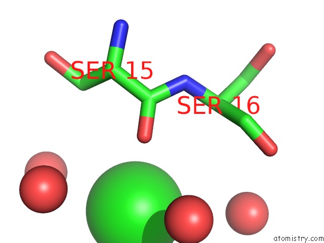
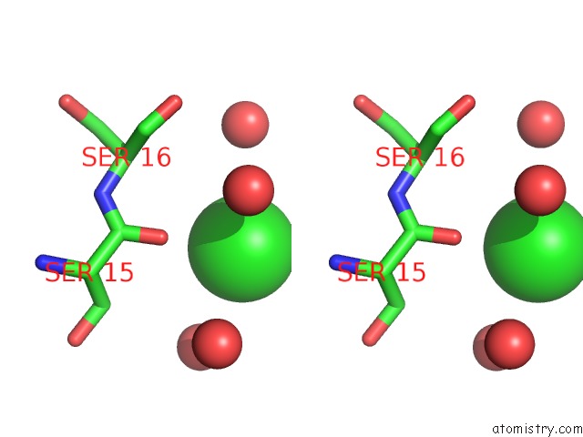
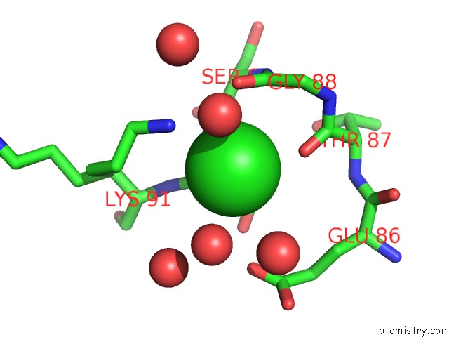
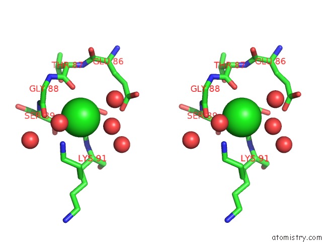
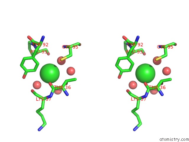
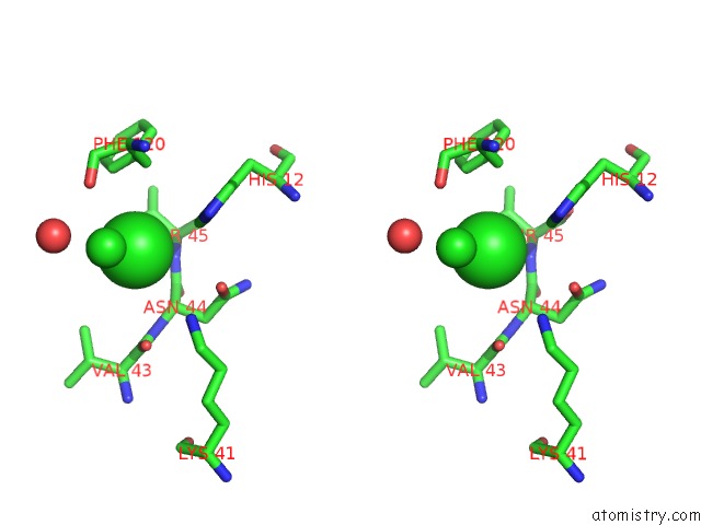
Chlorine binding site 1 out of 9 in 5na9
Go back to
Chlorine binding site 1 out
of 9 in the The X-Ray Structure of Bovine Pancreatic Ribonuclease Incubated in the Presence of An Excess of Carboplatin (1:10 Ratio)
Mono view
Stereo pair view
Mono view
Stereo pair view
A full contact list of Chlorine with other atoms in the Cl binding
site number 1 of The X-Ray Structure of Bovine Pancreatic Ribonuclease Incubated in the Presence of An Excess of Carboplatin (1:10 Ratio) within 5.0Å range:
|
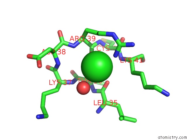
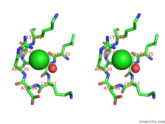
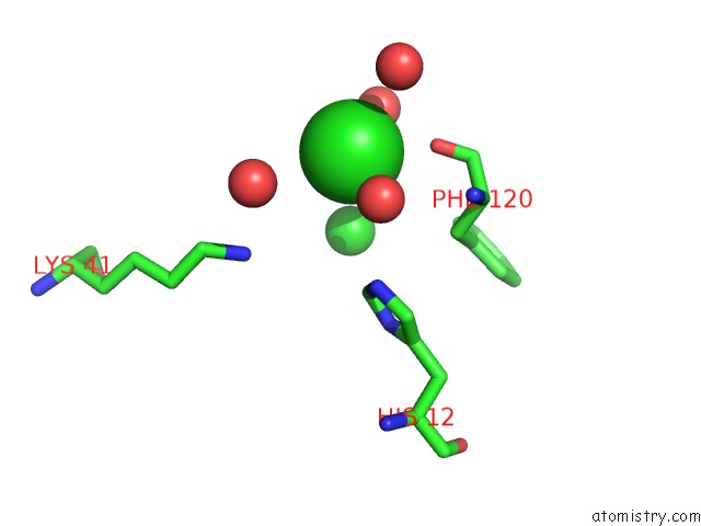
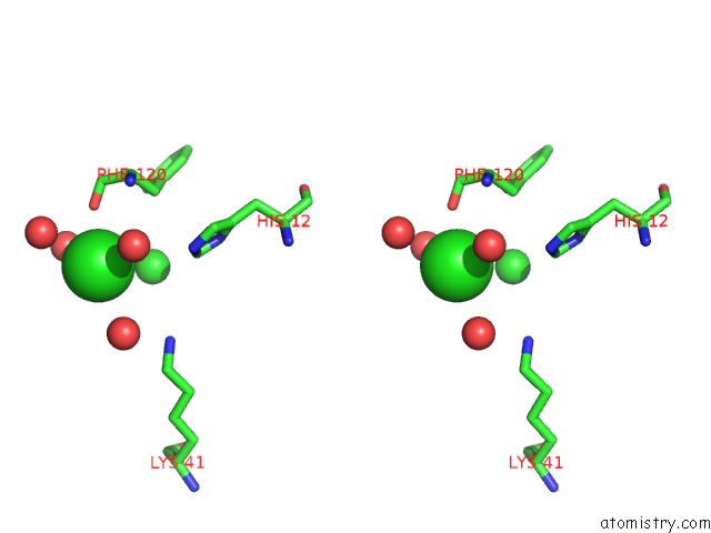
Chlorine binding site 2 out of 9 in 5na9
Go back to
Chlorine binding site 2 out
of 9 in the The X-Ray Structure of Bovine Pancreatic Ribonuclease Incubated in the Presence of An Excess of Carboplatin (1:10 Ratio)
Mono view
Stereo pair view
Mono view
Stereo pair view
A full contact list of Chlorine with other atoms in the Cl binding
site number 2 of The X-Ray Structure of Bovine Pancreatic Ribonuclease Incubated in the Presence of An Excess of Carboplatin (1:10 Ratio) within 5.0Å range:
|
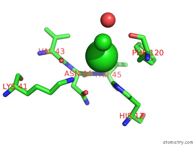
Chlorine binding site 3 out of 9 in 5na9
Go back to
Chlorine binding site 3 out
of 9 in the The X-Ray Structure of Bovine Pancreatic Ribonuclease Incubated in the Presence of An Excess of Carboplatin (1:10 Ratio)
Mono view
Stereo pair view
Mono view
Stereo pair view
A full contact list of Chlorine with other atoms in the Cl binding
site number 3 of The X-Ray Structure of Bovine Pancreatic Ribonuclease Incubated in the Presence of An Excess of Carboplatin (1:10 Ratio) within 5.0Å range:
|
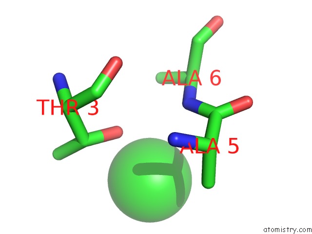
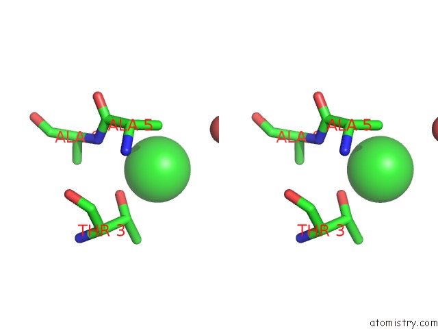
Chlorine binding site 4 out of 9 in 5na9
Go back to
Chlorine binding site 4 out
of 9 in the The X-Ray Structure of Bovine Pancreatic Ribonuclease Incubated in the Presence of An Excess of Carboplatin (1:10 Ratio)
Mono view
Stereo pair view
Mono view
Stereo pair view
A full contact list of Chlorine with other atoms in the Cl binding
site number 4 of The X-Ray Structure of Bovine Pancreatic Ribonuclease Incubated in the Presence of An Excess of Carboplatin (1:10 Ratio) within 5.0Å range:
|
Chlorine binding site 5 out of 9 in 5na9
Go back to
Chlorine binding site 5 out
of 9 in the The X-Ray Structure of Bovine Pancreatic Ribonuclease Incubated in the Presence of An Excess of Carboplatin (1:10 Ratio)
Mono view
Stereo pair view
Mono view
Stereo pair view
A full contact list of Chlorine with other atoms in the Cl binding
site number 5 of The X-Ray Structure of Bovine Pancreatic Ribonuclease Incubated in the Presence of An Excess of Carboplatin (1:10 Ratio) within 5.0Å range:
|
Chlorine binding site 6 out of 9 in 5na9
Go back to
Chlorine binding site 6 out
of 9 in the The X-Ray Structure of Bovine Pancreatic Ribonuclease Incubated in the Presence of An Excess of Carboplatin (1:10 Ratio)
Mono view
Stereo pair view
Mono view
Stereo pair view
A full contact list of Chlorine with other atoms in the Cl binding
site number 6 of The X-Ray Structure of Bovine Pancreatic Ribonuclease Incubated in the Presence of An Excess of Carboplatin (1:10 Ratio) within 5.0Å range:
|
Chlorine binding site 7 out of 9 in 5na9
Go back to
Chlorine binding site 7 out
of 9 in the The X-Ray Structure of Bovine Pancreatic Ribonuclease Incubated in the Presence of An Excess of Carboplatin (1:10 Ratio)

Mono view
Stereo pair view
Mono view
Stereo pair view
A full contact list of Chlorine with other atoms in the Cl binding
site number 7 of The X-Ray Structure of Bovine Pancreatic Ribonuclease Incubated in the Presence of An Excess of Carboplatin (1:10 Ratio) within 5.0Å range:
|
Chlorine binding site 8 out of 9 in 5na9
Go back to
Chlorine binding site 8 out
of 9 in the The X-Ray Structure of Bovine Pancreatic Ribonuclease Incubated in the Presence of An Excess of Carboplatin (1:10 Ratio)
Mono view
Stereo pair view
Mono view
Stereo pair view
A full contact list of Chlorine with other atoms in the Cl binding
site number 8 of The X-Ray Structure of Bovine Pancreatic Ribonuclease Incubated in the Presence of An Excess of Carboplatin (1:10 Ratio) within 5.0Å range:
|
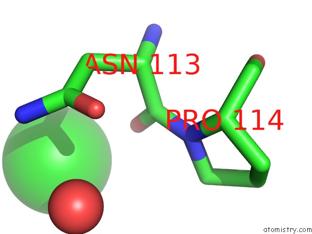
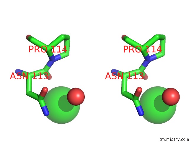
Chlorine binding site 9 out of 9 in 5na9
Go back to
Chlorine binding site 9 out
of 9 in the The X-Ray Structure of Bovine Pancreatic Ribonuclease Incubated in the Presence of An Excess of Carboplatin (1:10 Ratio)
Mono view
Stereo pair view
Mono view
Stereo pair view
A full contact list of Chlorine with other atoms in the Cl binding
site number 9 of The X-Ray Structure of Bovine Pancreatic Ribonuclease Incubated in the Presence of An Excess of Carboplatin (1:10 Ratio) within 5.0Å range:
|
Reference:
D.Picone,
F.Donnarumma,
G.Ferraro,
G.Gotte,
A.Fagagnini,
G.Butera,
M.Donadelli,
A.Merlino.
A Comparison Study on Rnase A Oligomerization Induced By Cisplatin, Carboplatin and Oxaliplatin. J. Inorg. Biochem. V. 173 105 2017.
ISSN: ISSN 1873-3344
PubMed: 28511060
DOI: 10.1016/J.JINORGBIO.2017.05.005
Page generated: Sat Jul 12 05:56:48 2025
ISSN: ISSN 1873-3344
PubMed: 28511060
DOI: 10.1016/J.JINORGBIO.2017.05.005
Last articles
Fe in 6MYPFe in 6MYO
Fe in 6MSN
Fe in 6MX5
Fe in 6MV0
Fe in 6MEV
Fe in 6MQ6
Fe in 6MQ1
Fe in 6MQ0
Fe in 6MPY