Chlorine »
PDB 5nrz-5nyw »
5ntu »
Chlorine in PDB 5ntu: Crystal Structure of Human Pro-Myostatin Precursor at 2.6 A Resolution
Protein crystallography data
The structure of Crystal Structure of Human Pro-Myostatin Precursor at 2.6 A Resolution, PDB code: 5ntu
was solved by
T.R.Cotton,
G.Fischer,
M.Hyvonen,
with X-Ray Crystallography technique. A brief refinement statistics is given in the table below:
| Resolution Low / High (Å) | 28.27 / 2.58 |
| Space group | C 1 2 1 |
| Cell size a, b, c (Å), α, β, γ (°) | 168.157, 36.301, 120.448, 90.00, 104.39, 90.00 |
| R / Rfree (%) | 21.5 / 26 |
Chlorine Binding Sites:
The binding sites of Chlorine atom in the Crystal Structure of Human Pro-Myostatin Precursor at 2.6 A Resolution
(pdb code 5ntu). This binding sites where shown within
5.0 Angstroms radius around Chlorine atom.
In total 2 binding sites of Chlorine where determined in the Crystal Structure of Human Pro-Myostatin Precursor at 2.6 A Resolution, PDB code: 5ntu:
Jump to Chlorine binding site number: 1; 2;
In total 2 binding sites of Chlorine where determined in the Crystal Structure of Human Pro-Myostatin Precursor at 2.6 A Resolution, PDB code: 5ntu:
Jump to Chlorine binding site number: 1; 2;
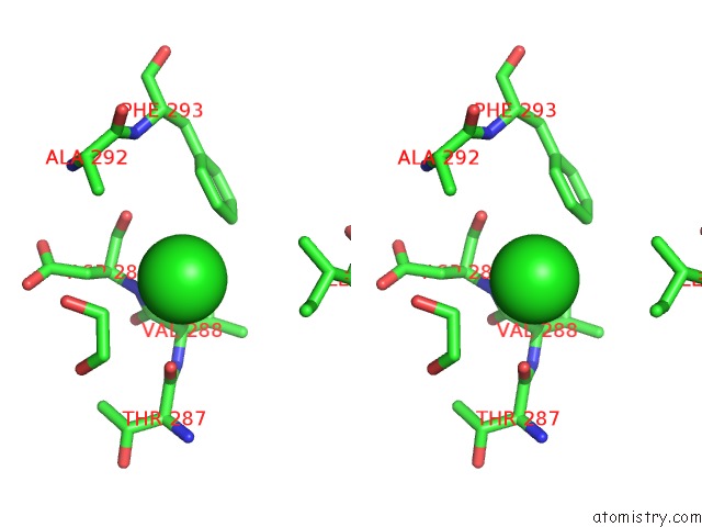
Chlorine binding site 1 out of 2 in 5ntu
Go back to
Chlorine binding site 1 out
of 2 in the Crystal Structure of Human Pro-Myostatin Precursor at 2.6 A Resolution

Mono view

Stereo pair view
Mono view
Stereo pair view
A full contact list of Chlorine with other atoms in the Cl binding
site number 1 of Crystal Structure of Human Pro-Myostatin Precursor at 2.6 A Resolution within 5.0Å range:
|
Chlorine binding site 2 out of 2 in 5ntu
Go back to
Chlorine binding site 2 out
of 2 in the Crystal Structure of Human Pro-Myostatin Precursor at 2.6 A Resolution

Mono view
Stereo pair view
Mono view
Stereo pair view
A full contact list of Chlorine with other atoms in the Cl binding
site number 2 of Crystal Structure of Human Pro-Myostatin Precursor at 2.6 A Resolution within 5.0Å range:
|
Reference:
T.R.Cotton,
G.Fischer,
X.Wang,
J.C.Mccoy,
M.Czepnik,
T.B.Thompson,
M.Hyvonen.
Structure of the Human Myostatin Precursor and Determinants of Growth Factor Latency. Embo J. V. 37 367 2018.
ISSN: ESSN 1460-2075
PubMed: 29330193
DOI: 10.15252/EMBJ.201797883
Page generated: Sat Jul 12 06:20:34 2025
ISSN: ESSN 1460-2075
PubMed: 29330193
DOI: 10.15252/EMBJ.201797883
Last articles
Na in 6N6DNa in 6N6C
Na in 6N6A
Na in 6MZ2
Na in 6N2V
Na in 6N2T
Na in 6N1C
Na in 6N2S
Na in 6MZ1
Na in 6N2R