Chlorine »
PDB 5o5u-5of0 »
5obh »
Chlorine in PDB 5obh: Crystal Structure of Glycine Binding Protein in Complex with Bicuculline
Protein crystallography data
The structure of Crystal Structure of Glycine Binding Protein in Complex with Bicuculline, PDB code: 5obh
was solved by
A.Dawson,
W.N.Hunter,
M.Jones,
with X-Ray Crystallography technique. A brief refinement statistics is given in the table below:
| Resolution Low / High (Å) | 46.82 / 2.40 |
| Space group | P 21 21 21 |
| Cell size a, b, c (Å), α, β, γ (°) | 71.243, 132.236, 132.631, 90.00, 90.00, 90.00 |
| R / Rfree (%) | 23 / 24.9 |
Chlorine Binding Sites:
The binding sites of Chlorine atom in the Crystal Structure of Glycine Binding Protein in Complex with Bicuculline
(pdb code 5obh). This binding sites where shown within
5.0 Angstroms radius around Chlorine atom.
In total 5 binding sites of Chlorine where determined in the Crystal Structure of Glycine Binding Protein in Complex with Bicuculline, PDB code: 5obh:
Jump to Chlorine binding site number: 1; 2; 3; 4; 5;
In total 5 binding sites of Chlorine where determined in the Crystal Structure of Glycine Binding Protein in Complex with Bicuculline, PDB code: 5obh:
Jump to Chlorine binding site number: 1; 2; 3; 4; 5;
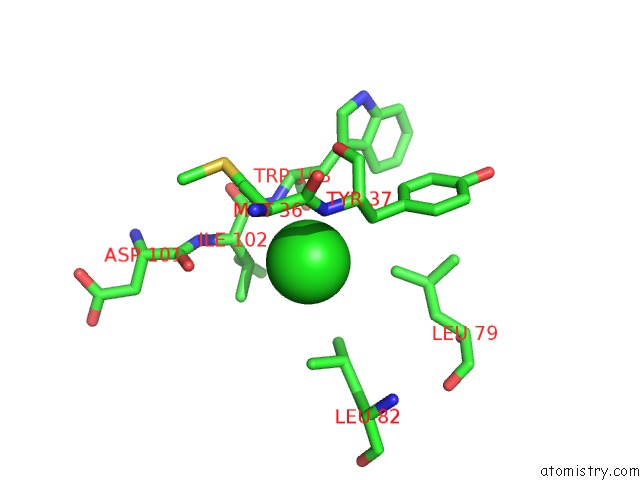
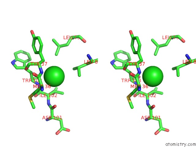
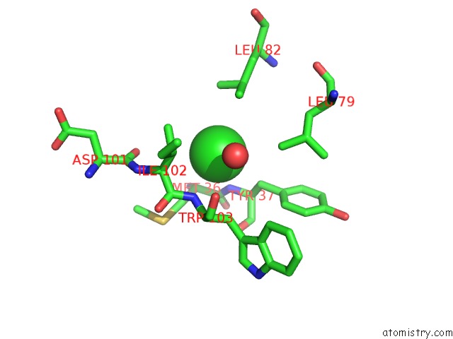
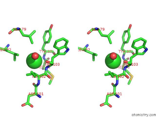
Chlorine binding site 1 out of 5 in 5obh
Go back to
Chlorine binding site 1 out
of 5 in the Crystal Structure of Glycine Binding Protein in Complex with Bicuculline
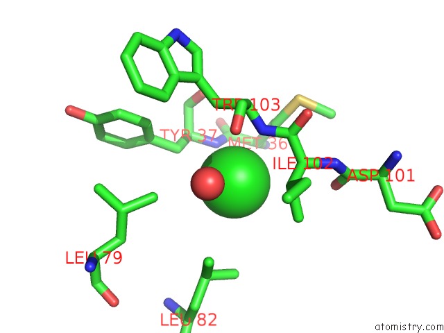
Mono view
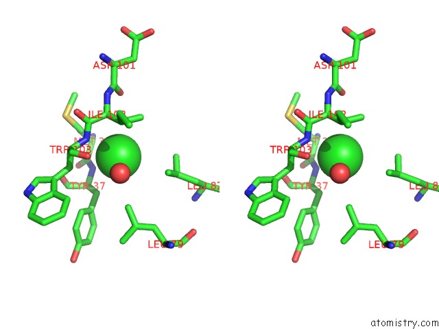
Stereo pair view
Mono view
Stereo pair view
A full contact list of Chlorine with other atoms in the Cl binding
site number 1 of Crystal Structure of Glycine Binding Protein in Complex with Bicuculline within 5.0Å range:
|
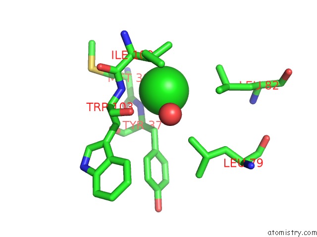
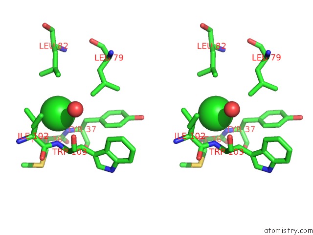
Chlorine binding site 2 out of 5 in 5obh
Go back to
Chlorine binding site 2 out
of 5 in the Crystal Structure of Glycine Binding Protein in Complex with Bicuculline
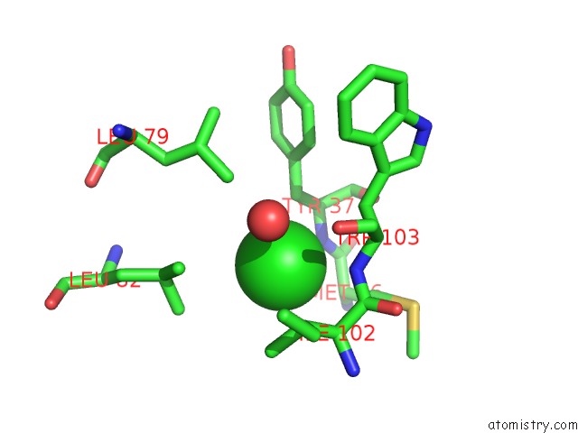
Mono view
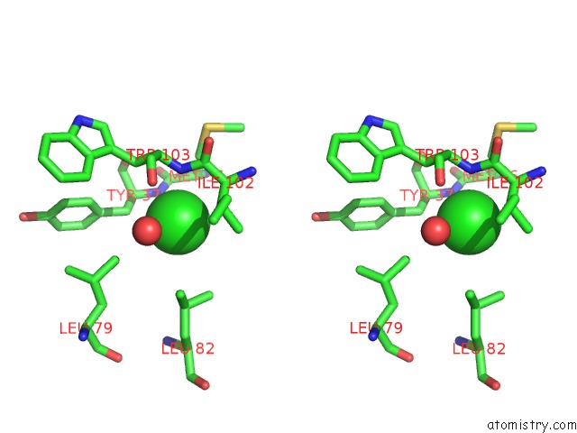
Stereo pair view
Mono view
Stereo pair view
A full contact list of Chlorine with other atoms in the Cl binding
site number 2 of Crystal Structure of Glycine Binding Protein in Complex with Bicuculline within 5.0Å range:
|
Chlorine binding site 3 out of 5 in 5obh
Go back to
Chlorine binding site 3 out
of 5 in the Crystal Structure of Glycine Binding Protein in Complex with Bicuculline
Mono view
Stereo pair view
Mono view
Stereo pair view
A full contact list of Chlorine with other atoms in the Cl binding
site number 3 of Crystal Structure of Glycine Binding Protein in Complex with Bicuculline within 5.0Å range:
|
Chlorine binding site 4 out of 5 in 5obh
Go back to
Chlorine binding site 4 out
of 5 in the Crystal Structure of Glycine Binding Protein in Complex with Bicuculline
Mono view
Stereo pair view
Mono view
Stereo pair view
A full contact list of Chlorine with other atoms in the Cl binding
site number 4 of Crystal Structure of Glycine Binding Protein in Complex with Bicuculline within 5.0Å range:
|
Chlorine binding site 5 out of 5 in 5obh
Go back to
Chlorine binding site 5 out
of 5 in the Crystal Structure of Glycine Binding Protein in Complex with Bicuculline
Mono view
Stereo pair view
Mono view
Stereo pair view
A full contact list of Chlorine with other atoms in the Cl binding
site number 5 of Crystal Structure of Glycine Binding Protein in Complex with Bicuculline within 5.0Å range:
|
Reference:
A.Dawson,
W.N.Hunter,
M.Jones.
Crystal Structure of Glycine Binding Protein in Complex with Bicuculline To Be Published.
Page generated: Sat Jul 12 06:40:53 2025
Last articles
Mg in 4JJSMg in 4JJ2
Mg in 4JIW
Mg in 4JIV
Mg in 4JIB
Mg in 4JI4
Mg in 4JI5
Mg in 4JI1
Mg in 4JI0
Mg in 4JI2