Chlorine »
PDB 5o5u-5of0 »
5odj »
Chlorine in PDB 5odj: Single-Stranded Dna-Binding Protein From Bacteriophage ENC34
Protein crystallography data
The structure of Single-Stranded Dna-Binding Protein From Bacteriophage ENC34, PDB code: 5odj
was solved by
E.Cernooka,
J.Rumnieks,
A.Kazaks,
K.Tars,
with X-Ray Crystallography technique. A brief refinement statistics is given in the table below:
| Resolution Low / High (Å) | 30.78 / 1.50 |
| Space group | P 21 21 21 |
| Cell size a, b, c (Å), α, β, γ (°) | 38.080, 67.200, 76.830, 90.00, 90.00, 90.00 |
| R / Rfree (%) | 15.7 / 19.3 |
Other elements in 5odj:
The structure of Single-Stranded Dna-Binding Protein From Bacteriophage ENC34 also contains other interesting chemical elements:
| Magnesium | (Mg) | 1 atom |
Chlorine Binding Sites:
The binding sites of Chlorine atom in the Single-Stranded Dna-Binding Protein From Bacteriophage ENC34
(pdb code 5odj). This binding sites where shown within
5.0 Angstroms radius around Chlorine atom.
In total only one binding site of Chlorine was determined in the Single-Stranded Dna-Binding Protein From Bacteriophage ENC34, PDB code: 5odj:
In total only one binding site of Chlorine was determined in the Single-Stranded Dna-Binding Protein From Bacteriophage ENC34, PDB code: 5odj:
Chlorine binding site 1 out of 1 in 5odj
Go back to
Chlorine binding site 1 out
of 1 in the Single-Stranded Dna-Binding Protein From Bacteriophage ENC34
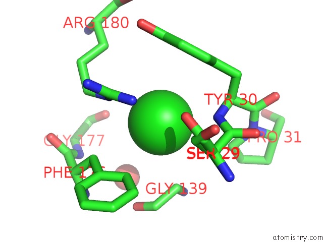
Mono view
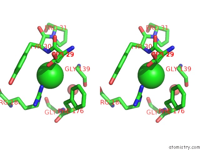
Stereo pair view
Mono view
Stereo pair view
A full contact list of Chlorine with other atoms in the Cl binding
site number 1 of Single-Stranded Dna-Binding Protein From Bacteriophage ENC34 within 5.0Å range:
|
Reference:
E.Cernooka,
J.Rumnieks,
K.Tars,
A.Kazaks.
Structural Basis For Dna Recognition of A Single-Stranded Dna-Binding Protein From Enterobacter Phage ENC34. Sci Rep V. 7 15529 2017.
ISSN: ESSN 2045-2322
PubMed: 29138440
DOI: 10.1038/S41598-017-15774-Y
Page generated: Sat Jul 12 06:42:27 2025
ISSN: ESSN 2045-2322
PubMed: 29138440
DOI: 10.1038/S41598-017-15774-Y
Last articles
Mg in 5MRAMg in 5MTV
Mg in 5MS0
Mg in 5MRU
Mg in 5MQJ
Mg in 5MQW
Mg in 5MQT
Mg in 5MQL
Mg in 5MQ1
Mg in 5MQ0