Chlorine »
PDB 5qa7-5qcc »
5qby »
Chlorine in PDB 5qby: Crystal Structure of Human Cathepsin-S with Bound Ligand
Enzymatic activity of Crystal Structure of Human Cathepsin-S with Bound Ligand
All present enzymatic activity of Crystal Structure of Human Cathepsin-S with Bound Ligand:
3.4.22.27;
3.4.22.27;
Protein crystallography data
The structure of Crystal Structure of Human Cathepsin-S with Bound Ligand, PDB code: 5qby
was solved by
S.D.Bembenek,
M.K.Ameriks,
T.Mirzadegan,
H.Yang,
C.Shao,
S.K.Burley,
with X-Ray Crystallography technique. A brief refinement statistics is given in the table below:
| Resolution Low / High (Å) | 33.87 / 2.25 |
| Space group | C 1 2 1 |
| Cell size a, b, c (Å), α, β, γ (°) | 171.238, 34.376, 103.815, 90.00, 125.10, 90.00 |
| R / Rfree (%) | 17.1 / 20.4 |
Other elements in 5qby:
The structure of Crystal Structure of Human Cathepsin-S with Bound Ligand also contains other interesting chemical elements:
| Fluorine | (F) | 2 atoms |
Chlorine Binding Sites:
The binding sites of Chlorine atom in the Crystal Structure of Human Cathepsin-S with Bound Ligand
(pdb code 5qby). This binding sites where shown within
5.0 Angstroms radius around Chlorine atom.
In total 4 binding sites of Chlorine where determined in the Crystal Structure of Human Cathepsin-S with Bound Ligand, PDB code: 5qby:
Jump to Chlorine binding site number: 1; 2; 3; 4;
In total 4 binding sites of Chlorine where determined in the Crystal Structure of Human Cathepsin-S with Bound Ligand, PDB code: 5qby:
Jump to Chlorine binding site number: 1; 2; 3; 4;
Chlorine binding site 1 out of 4 in 5qby
Go back to
Chlorine binding site 1 out
of 4 in the Crystal Structure of Human Cathepsin-S with Bound Ligand

Mono view
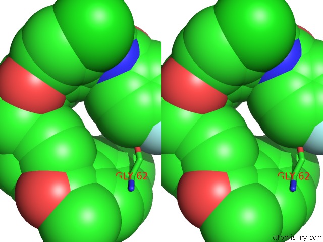
Stereo pair view
Mono view
Stereo pair view
A full contact list of Chlorine with other atoms in the Cl binding
site number 1 of Crystal Structure of Human Cathepsin-S with Bound Ligand within 5.0Å range:
|
Chlorine binding site 2 out of 4 in 5qby
Go back to
Chlorine binding site 2 out
of 4 in the Crystal Structure of Human Cathepsin-S with Bound Ligand
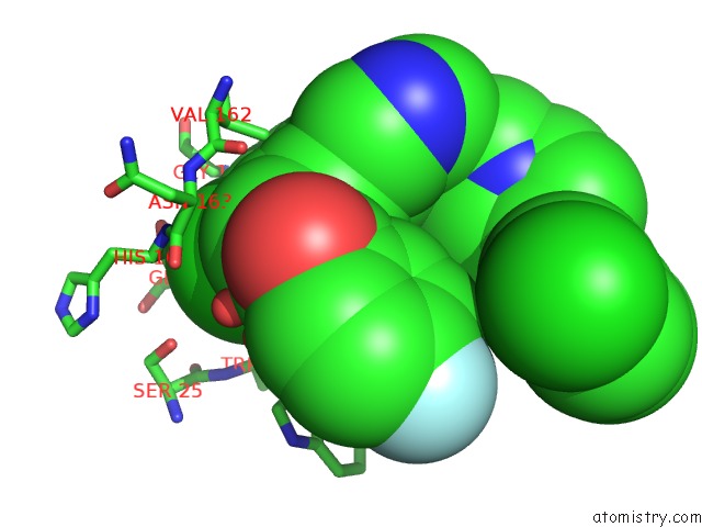
Mono view
Stereo pair view
Mono view
Stereo pair view
A full contact list of Chlorine with other atoms in the Cl binding
site number 2 of Crystal Structure of Human Cathepsin-S with Bound Ligand within 5.0Å range:
|
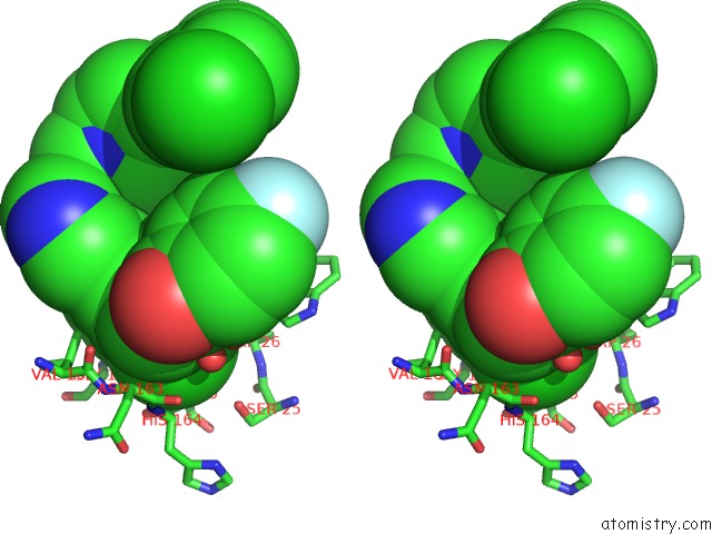
Chlorine binding site 3 out of 4 in 5qby
Go back to
Chlorine binding site 3 out
of 4 in the Crystal Structure of Human Cathepsin-S with Bound Ligand

Mono view

Stereo pair view
Mono view
Stereo pair view
A full contact list of Chlorine with other atoms in the Cl binding
site number 3 of Crystal Structure of Human Cathepsin-S with Bound Ligand within 5.0Å range:
|
Chlorine binding site 4 out of 4 in 5qby
Go back to
Chlorine binding site 4 out
of 4 in the Crystal Structure of Human Cathepsin-S with Bound Ligand
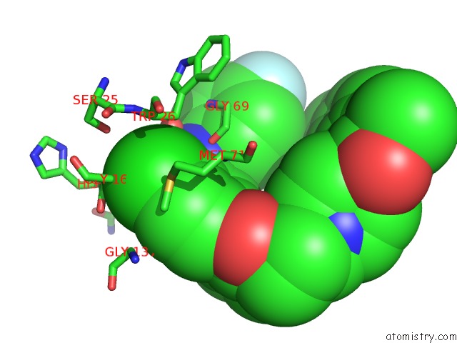
Mono view

Stereo pair view
Mono view
Stereo pair view
A full contact list of Chlorine with other atoms in the Cl binding
site number 4 of Crystal Structure of Human Cathepsin-S with Bound Ligand within 5.0Å range:
|
Reference:
M.K.Ameriks,
S.D.Bembenek,
M.T.Burdett,
I.C.Choong,
J.P.Edwards,
D.Gebauer,
Y.Gu,
L.Karlsson,
H.E.Purkey,
B.L.Staker,
S.Sun,
R.L.Thurmond,
J.Zhu.
Diazinones As P2 Replacements For Pyrazole-Based Cathepsin S Inhibitors Bioorg.Med.Chem.Lett. V. 20 4060 2010.
ISSN: ISSN 0960-894X
PubMed: 20541404
DOI: 10.1016/J.BMCL.2010.05.086
Page generated: Sat Jul 12 07:25:36 2025
ISSN: ISSN 0960-894X
PubMed: 20541404
DOI: 10.1016/J.BMCL.2010.05.086
Last articles
I in 8BIRI in 8C2G
I in 8BW9
I in 8BVX
I in 8ATE
I in 8AIL
I in 8A5Z
I in 8A56
I in 8A31
I in 8A32