Chlorine »
PDB 5siu-5spo »
5sk9 »
Chlorine in PDB 5sk9: Crystal Structure of Human Phosphodiesterase 10 in Complex with 2- Chloro-5-Methoxy-3-Methylquinoxaline
Enzymatic activity of Crystal Structure of Human Phosphodiesterase 10 in Complex with 2- Chloro-5-Methoxy-3-Methylquinoxaline
All present enzymatic activity of Crystal Structure of Human Phosphodiesterase 10 in Complex with 2- Chloro-5-Methoxy-3-Methylquinoxaline:
3.1.4.17;
3.1.4.17;
Protein crystallography data
The structure of Crystal Structure of Human Phosphodiesterase 10 in Complex with 2- Chloro-5-Methoxy-3-Methylquinoxaline, PDB code: 5sk9
was solved by
C.Joseph,
J.Benz,
A.Flohr,
M.Boehringer,
M.G.Rudolph,
with X-Ray Crystallography technique. A brief refinement statistics is given in the table below:
| Resolution Low / High (Å) | 43.64 / 2.82 |
| Space group | H 3 |
| Cell size a, b, c (Å), α, β, γ (°) | 135.698, 135.698, 234.384, 90, 90, 120 |
| R / Rfree (%) | 21.2 / 23.9 |
Other elements in 5sk9:
The structure of Crystal Structure of Human Phosphodiesterase 10 in Complex with 2- Chloro-5-Methoxy-3-Methylquinoxaline also contains other interesting chemical elements:
| Zinc | (Zn) | 4 atoms |
| Magnesium | (Mg) | 4 atoms |
Chlorine Binding Sites:
The binding sites of Chlorine atom in the Crystal Structure of Human Phosphodiesterase 10 in Complex with 2- Chloro-5-Methoxy-3-Methylquinoxaline
(pdb code 5sk9). This binding sites where shown within
5.0 Angstroms radius around Chlorine atom.
In total 4 binding sites of Chlorine where determined in the Crystal Structure of Human Phosphodiesterase 10 in Complex with 2- Chloro-5-Methoxy-3-Methylquinoxaline, PDB code: 5sk9:
Jump to Chlorine binding site number: 1; 2; 3; 4;
In total 4 binding sites of Chlorine where determined in the Crystal Structure of Human Phosphodiesterase 10 in Complex with 2- Chloro-5-Methoxy-3-Methylquinoxaline, PDB code: 5sk9:
Jump to Chlorine binding site number: 1; 2; 3; 4;
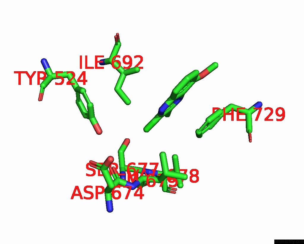
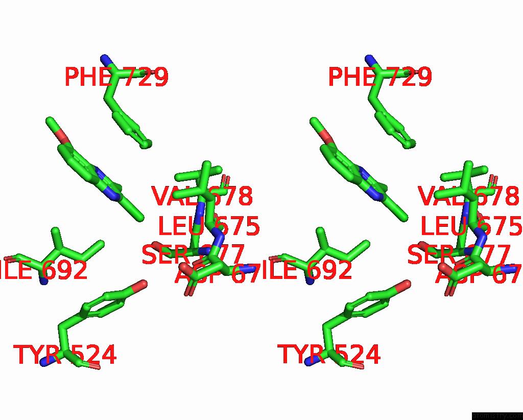
Chlorine binding site 1 out of 4 in 5sk9
Go back to
Chlorine binding site 1 out
of 4 in the Crystal Structure of Human Phosphodiesterase 10 in Complex with 2- Chloro-5-Methoxy-3-Methylquinoxaline
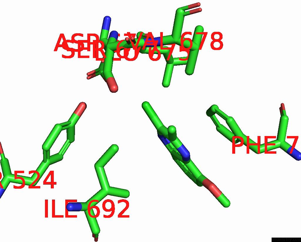
Mono view
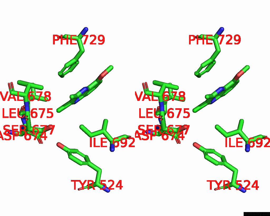
Stereo pair view
Mono view
Stereo pair view
A full contact list of Chlorine with other atoms in the Cl binding
site number 1 of Crystal Structure of Human Phosphodiesterase 10 in Complex with 2- Chloro-5-Methoxy-3-Methylquinoxaline within 5.0Å range:
|
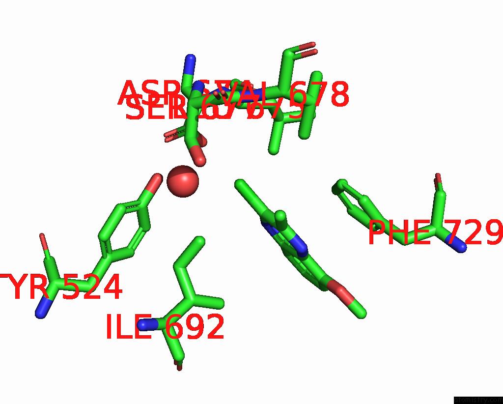
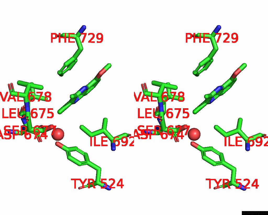
Chlorine binding site 2 out of 4 in 5sk9
Go back to
Chlorine binding site 2 out
of 4 in the Crystal Structure of Human Phosphodiesterase 10 in Complex with 2- Chloro-5-Methoxy-3-Methylquinoxaline
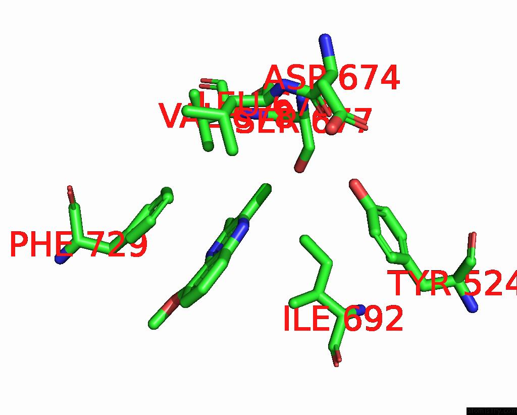
Mono view
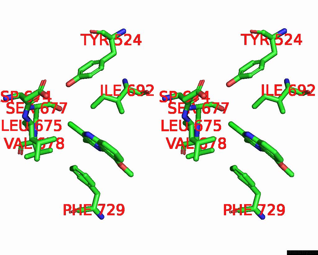
Stereo pair view
Mono view
Stereo pair view
A full contact list of Chlorine with other atoms in the Cl binding
site number 2 of Crystal Structure of Human Phosphodiesterase 10 in Complex with 2- Chloro-5-Methoxy-3-Methylquinoxaline within 5.0Å range:
|
Chlorine binding site 3 out of 4 in 5sk9
Go back to
Chlorine binding site 3 out
of 4 in the Crystal Structure of Human Phosphodiesterase 10 in Complex with 2- Chloro-5-Methoxy-3-Methylquinoxaline
Mono view
Stereo pair view
Mono view
Stereo pair view
A full contact list of Chlorine with other atoms in the Cl binding
site number 3 of Crystal Structure of Human Phosphodiesterase 10 in Complex with 2- Chloro-5-Methoxy-3-Methylquinoxaline within 5.0Å range:
|
Chlorine binding site 4 out of 4 in 5sk9
Go back to
Chlorine binding site 4 out
of 4 in the Crystal Structure of Human Phosphodiesterase 10 in Complex with 2- Chloro-5-Methoxy-3-Methylquinoxaline
Mono view
Stereo pair view
Mono view
Stereo pair view
A full contact list of Chlorine with other atoms in the Cl binding
site number 4 of Crystal Structure of Human Phosphodiesterase 10 in Complex with 2- Chloro-5-Methoxy-3-Methylquinoxaline within 5.0Å range:
|
Reference:
A.Flohr,
D.Schlatter,
B.Kuhn,
M.G.Rudolph.
Crystal Structure of A Human Phosphodiesterase 10 Complex To Be Published.
Page generated: Sat Jul 12 08:34:14 2025
Last articles
Mg in 7M44Mg in 7M2M
Mg in 7M1T
Mg in 7M13
Mg in 7M1Q
Mg in 7M0Z
Mg in 7M0Y
Mg in 7M0X
Mg in 7M0W
Mg in 7M0E