Chlorine »
PDB 5tic-5trc »
5tjy »
Chlorine in PDB 5tjy: Structure of 4-Hydroxy-Tetrahydrodipicolinate Reductase From Mycobacterium Tuberculosis with 2,6 Pyridine Dicarboxylic Acid and Nadh
Enzymatic activity of Structure of 4-Hydroxy-Tetrahydrodipicolinate Reductase From Mycobacterium Tuberculosis with 2,6 Pyridine Dicarboxylic Acid and Nadh
All present enzymatic activity of Structure of 4-Hydroxy-Tetrahydrodipicolinate Reductase From Mycobacterium Tuberculosis with 2,6 Pyridine Dicarboxylic Acid and Nadh:
1.17.1.8;
1.17.1.8;
Protein crystallography data
The structure of Structure of 4-Hydroxy-Tetrahydrodipicolinate Reductase From Mycobacterium Tuberculosis with 2,6 Pyridine Dicarboxylic Acid and Nadh, PDB code: 5tjy
was solved by
N.Mank,
K.Arnette,
V.Klapper,
M.Chruszcz,
with X-Ray Crystallography technique. A brief refinement statistics is given in the table below:
| Resolution Low / High (Å) | 40.00 / 2.40 |
| Space group | P 62 2 2 |
| Cell size a, b, c (Å), α, β, γ (°) | 66.421, 66.421, 249.363, 90.00, 90.00, 120.00 |
| R / Rfree (%) | 14.6 / 19.5 |
Other elements in 5tjy:
The structure of Structure of 4-Hydroxy-Tetrahydrodipicolinate Reductase From Mycobacterium Tuberculosis with 2,6 Pyridine Dicarboxylic Acid and Nadh also contains other interesting chemical elements:
| Sodium | (Na) | 1 atom |
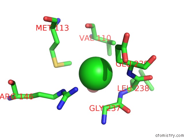
Chlorine Binding Sites:
The binding sites of Chlorine atom in the Structure of 4-Hydroxy-Tetrahydrodipicolinate Reductase From Mycobacterium Tuberculosis with 2,6 Pyridine Dicarboxylic Acid and Nadh
(pdb code 5tjy). This binding sites where shown within
5.0 Angstroms radius around Chlorine atom.
In total only one binding site of Chlorine was determined in the Structure of 4-Hydroxy-Tetrahydrodipicolinate Reductase From Mycobacterium Tuberculosis with 2,6 Pyridine Dicarboxylic Acid and Nadh, PDB code: 5tjy:
In total only one binding site of Chlorine was determined in the Structure of 4-Hydroxy-Tetrahydrodipicolinate Reductase From Mycobacterium Tuberculosis with 2,6 Pyridine Dicarboxylic Acid and Nadh, PDB code: 5tjy:
Chlorine binding site 1 out of 1 in 5tjy
Go back to
Chlorine binding site 1 out
of 1 in the Structure of 4-Hydroxy-Tetrahydrodipicolinate Reductase From Mycobacterium Tuberculosis with 2,6 Pyridine Dicarboxylic Acid and Nadh
Mono view

Stereo pair view
Mono view
Stereo pair view
A full contact list of Chlorine with other atoms in the Cl binding
site number 1 of Structure of 4-Hydroxy-Tetrahydrodipicolinate Reductase From Mycobacterium Tuberculosis with 2,6 Pyridine Dicarboxylic Acid and Nadh within 5.0Å range:
|
Reference:
N.Mank,
K.Arnette,
V.Klapper,
M.Chruszcz.
Structure of 4-Hydroxy-Tetrahydrodipicolinate Reductase From Mycobacterium Tuberculosis with 2,6 Pyridine Dicarboxylic Acid and Nadh To Be Published.
Page generated: Sat Jul 12 08:57:24 2025
Last articles
Na in 1PA6Na in 1PH2
Na in 1P9E
Na in 1P59
Na in 1P4Y
Na in 1P4Z
Na in 1P54
Na in 1P2H
Na in 1P2E
Na in 1P0S