Chlorine »
PDB 5u0l-5u72 »
5u1o »
Chlorine in PDB 5u1o: 2.3 Angstrom Resolution Crystal Structure of Glutathione Reductase From Vibrio Parahaemolyticus in Complex with Fad.
Protein crystallography data
The structure of 2.3 Angstrom Resolution Crystal Structure of Glutathione Reductase From Vibrio Parahaemolyticus in Complex with Fad., PDB code: 5u1o
was solved by
G.Minasov,
L.Shuvalova,
A.Cardona-Correa,
I.Dubrovska,
S.Grimshaw,
K.Kwon,
W.F.Anderson,
Center For Structural Genomics Of Infectious Diseases(Csgid),
with X-Ray Crystallography technique. A brief refinement statistics is given in the table below:
| Resolution Low / High (Å) | 29.95 / 2.31 |
| Space group | P 21 21 21 |
| Cell size a, b, c (Å), α, β, γ (°) | 58.528, 174.289, 206.094, 90.00, 90.00, 90.00 |
| R / Rfree (%) | 20.2 / 24.9 |
Chlorine Binding Sites:
The binding sites of Chlorine atom in the 2.3 Angstrom Resolution Crystal Structure of Glutathione Reductase From Vibrio Parahaemolyticus in Complex with Fad.
(pdb code 5u1o). This binding sites where shown within
5.0 Angstroms radius around Chlorine atom.
In total only one binding site of Chlorine was determined in the 2.3 Angstrom Resolution Crystal Structure of Glutathione Reductase From Vibrio Parahaemolyticus in Complex with Fad., PDB code: 5u1o:
In total only one binding site of Chlorine was determined in the 2.3 Angstrom Resolution Crystal Structure of Glutathione Reductase From Vibrio Parahaemolyticus in Complex with Fad., PDB code: 5u1o:
Chlorine binding site 1 out of 1 in 5u1o
Go back to
Chlorine binding site 1 out
of 1 in the 2.3 Angstrom Resolution Crystal Structure of Glutathione Reductase From Vibrio Parahaemolyticus in Complex with Fad.
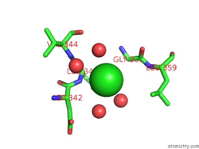
Mono view
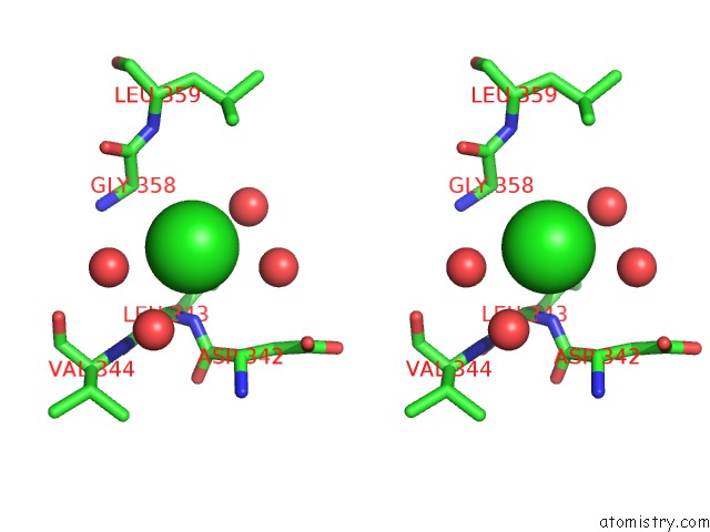
Stereo pair view
Mono view
Stereo pair view
A full contact list of Chlorine with other atoms in the Cl binding
site number 1 of 2.3 Angstrom Resolution Crystal Structure of Glutathione Reductase From Vibrio Parahaemolyticus in Complex with Fad. within 5.0Å range:
|
Reference:
G.Minasov,
L.Shuvalova,
A.Cardona-Correa,
I.Dubrovska,
S.Grimshaw,
K.Kwon,
W.F.Anderson,
Center For Structural Genomics Of Infectious Diseases(Csgid).
2.3 Angstrom Resolution Crystal Structure of Glutathione Reductase From Vibrio Parahaemolyticus in Complex with Fad. To Be Published.
Page generated: Sat Jul 12 09:11:21 2025
Last articles
Fe in 8DVYFe in 8DVP
Fe in 8DPN
Fe in 8DQV
Fe in 8DVX
Fe in 8DSG
Fe in 8DNP
Fe in 8DRM
Fe in 8DRL
Fe in 8DRJ