Chlorine »
PDB 5u0l-5u72 »
5u3q »
Chlorine in PDB 5u3q: Human Ppardelta Ligand-Binding Domain in Complexed with Specific Agonist 1
Protein crystallography data
The structure of Human Ppardelta Ligand-Binding Domain in Complexed with Specific Agonist 1, PDB code: 5u3q
was solved by
C.-C.Wu,
T.J.Baiga,
M.Downes,
J.J.La Clair,
A.R.Atkins,
S.B.Richard,
T.A.Stockley-Noel,
M.E.Bowman,
R.M.Evans,
J.P.Noel,
with X-Ray Crystallography technique. A brief refinement statistics is given in the table below:
| Resolution Low / High (Å) | 47.85 / 1.50 |
| Space group | P 1 21 1 |
| Cell size a, b, c (Å), α, β, γ (°) | 39.460, 94.350, 96.730, 90.00, 98.36, 90.00 |
| R / Rfree (%) | 15.9 / 18.9 |
Other elements in 5u3q:
The structure of Human Ppardelta Ligand-Binding Domain in Complexed with Specific Agonist 1 also contains other interesting chemical elements:
| Potassium | (K) | 2 atoms |
Chlorine Binding Sites:
The binding sites of Chlorine atom in the Human Ppardelta Ligand-Binding Domain in Complexed with Specific Agonist 1
(pdb code 5u3q). This binding sites where shown within
5.0 Angstroms radius around Chlorine atom.
In total 2 binding sites of Chlorine where determined in the Human Ppardelta Ligand-Binding Domain in Complexed with Specific Agonist 1, PDB code: 5u3q:
Jump to Chlorine binding site number: 1; 2;
In total 2 binding sites of Chlorine where determined in the Human Ppardelta Ligand-Binding Domain in Complexed with Specific Agonist 1, PDB code: 5u3q:
Jump to Chlorine binding site number: 1; 2;
Chlorine binding site 1 out of 2 in 5u3q
Go back to
Chlorine binding site 1 out
of 2 in the Human Ppardelta Ligand-Binding Domain in Complexed with Specific Agonist 1
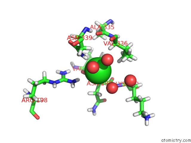
Mono view
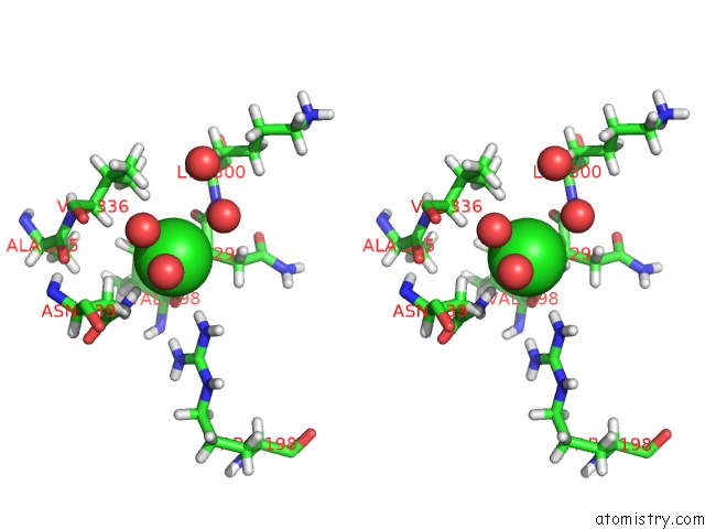
Stereo pair view
Mono view
Stereo pair view
A full contact list of Chlorine with other atoms in the Cl binding
site number 1 of Human Ppardelta Ligand-Binding Domain in Complexed with Specific Agonist 1 within 5.0Å range:
|
Chlorine binding site 2 out of 2 in 5u3q
Go back to
Chlorine binding site 2 out
of 2 in the Human Ppardelta Ligand-Binding Domain in Complexed with Specific Agonist 1
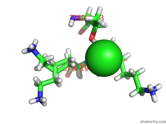
Mono view
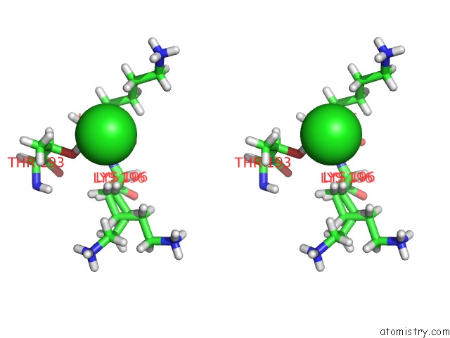
Stereo pair view
Mono view
Stereo pair view
A full contact list of Chlorine with other atoms in the Cl binding
site number 2 of Human Ppardelta Ligand-Binding Domain in Complexed with Specific Agonist 1 within 5.0Å range:
|
Reference:
C.C.Wu,
T.J.Baiga,
M.Downes,
J.J.La Clair,
A.R.Atkins,
S.B.Richard,
W.Fan,
T.A.Stockley-Noel,
M.E.Bowman,
J.P.Noel,
R.M.Evans.
Structural Basis For Specific Ligation of the Peroxisome Proliferator-Activated Receptor Delta. Proc. Natl. Acad. Sci. V. 114 E2563 2017U.S.A..
ISSN: ESSN 1091-6490
PubMed: 28320959
DOI: 10.1073/PNAS.1621513114
Page generated: Sat Jul 12 09:13:36 2025
ISSN: ESSN 1091-6490
PubMed: 28320959
DOI: 10.1073/PNAS.1621513114
Last articles
Fe in 8DVYFe in 8DVP
Fe in 8DPN
Fe in 8DQV
Fe in 8DVX
Fe in 8DSG
Fe in 8DNP
Fe in 8DRM
Fe in 8DRL
Fe in 8DRJ