Chlorine »
PDB 5u7k-5ufo »
5ucv »
Chlorine in PDB 5ucv: Crystal Structure of A Ribosome Biogenesis Gtp-Binding Protein (Ysxc) From Neisseria Gonorrhoeae with Bound Gdp
Protein crystallography data
The structure of Crystal Structure of A Ribosome Biogenesis Gtp-Binding Protein (Ysxc) From Neisseria Gonorrhoeae with Bound Gdp, PDB code: 5ucv
was solved by
Seattle Structural Genomics Center For Infectious Disease,
Seattlestructural Genomics Center For Infectious Disease (Ssgcid),
with X-Ray Crystallography technique. A brief refinement statistics is given in the table below:
| Resolution Low / High (Å) | 48.20 / 1.80 |
| Space group | P 21 21 21 |
| Cell size a, b, c (Å), α, β, γ (°) | 52.110, 68.440, 126.890, 90.00, 90.00, 90.00 |
| R / Rfree (%) | 16.7 / 20.7 |
Chlorine Binding Sites:
The binding sites of Chlorine atom in the Crystal Structure of A Ribosome Biogenesis Gtp-Binding Protein (Ysxc) From Neisseria Gonorrhoeae with Bound Gdp
(pdb code 5ucv). This binding sites where shown within
5.0 Angstroms radius around Chlorine atom.
In total only one binding site of Chlorine was determined in the Crystal Structure of A Ribosome Biogenesis Gtp-Binding Protein (Ysxc) From Neisseria Gonorrhoeae with Bound Gdp, PDB code: 5ucv:
In total only one binding site of Chlorine was determined in the Crystal Structure of A Ribosome Biogenesis Gtp-Binding Protein (Ysxc) From Neisseria Gonorrhoeae with Bound Gdp, PDB code: 5ucv:
Chlorine binding site 1 out of 1 in 5ucv
Go back to
Chlorine binding site 1 out
of 1 in the Crystal Structure of A Ribosome Biogenesis Gtp-Binding Protein (Ysxc) From Neisseria Gonorrhoeae with Bound Gdp
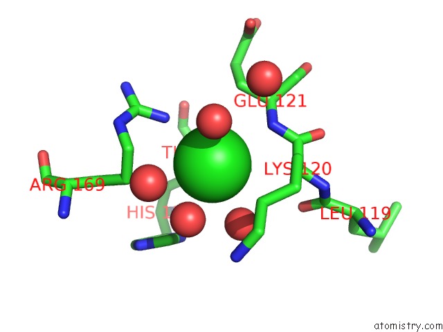
Mono view
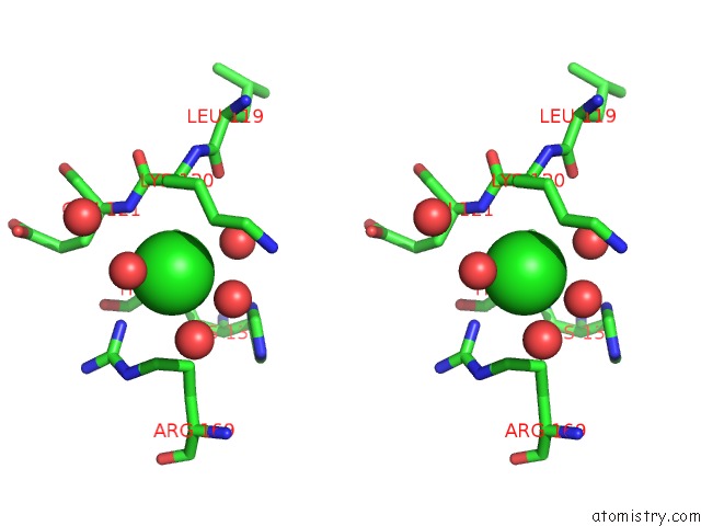
Stereo pair view
Mono view
Stereo pair view
A full contact list of Chlorine with other atoms in the Cl binding
site number 1 of Crystal Structure of A Ribosome Biogenesis Gtp-Binding Protein (Ysxc) From Neisseria Gonorrhoeae with Bound Gdp within 5.0Å range:
|
Reference:
D.M.Dranow,
S.J.Mayclin,
D.D.Lorimer,
T.E.Edwards.
Crystal Structure of A Ribosome Biogenesis Gtp-Binding Protein (Ysxc) From Neisseria Gonorrhoeae with Bound Gdp To Be Published.
Page generated: Sat Jul 12 09:18:53 2025
Last articles
Mn in 8D3QMn in 8D3L
Mn in 8D3P
Mn in 8D3M
Mn in 8CTF
Mn in 8BRP
Mn in 8C4O
Mn in 8CRD
Mn in 8COH
Mn in 8C7U