Chlorine »
PDB 5ufq-5up7 »
5ujs »
Chlorine in PDB 5ujs: 2.45 Angstrom Resolution Crystal Structure of Udp-N-Acetylglucosamine 1-Carboxyvinyltransferase From Campylobacter Jejuni.
Enzymatic activity of 2.45 Angstrom Resolution Crystal Structure of Udp-N-Acetylglucosamine 1-Carboxyvinyltransferase From Campylobacter Jejuni.
All present enzymatic activity of 2.45 Angstrom Resolution Crystal Structure of Udp-N-Acetylglucosamine 1-Carboxyvinyltransferase From Campylobacter Jejuni.:
2.5.1.7;
2.5.1.7;
Protein crystallography data
The structure of 2.45 Angstrom Resolution Crystal Structure of Udp-N-Acetylglucosamine 1-Carboxyvinyltransferase From Campylobacter Jejuni., PDB code: 5ujs
was solved by
G.Minasov,
L.Shuvalova,
I.Dubrovska,
J.Winsor,
J.Stam,
K.Kwon,
W.F.Anderson,
Center For Structural Genomics Of Infectious Diseases(Csgid),
with X-Ray Crystallography technique. A brief refinement statistics is given in the table below:
| Resolution Low / High (Å) | 29.93 / 2.46 |
| Space group | C 1 2 1 |
| Cell size a, b, c (Å), α, β, γ (°) | 130.258, 86.024, 80.421, 90.00, 109.28, 90.00 |
| R / Rfree (%) | 22.4 / 27.5 |
Chlorine Binding Sites:
The binding sites of Chlorine atom in the 2.45 Angstrom Resolution Crystal Structure of Udp-N-Acetylglucosamine 1-Carboxyvinyltransferase From Campylobacter Jejuni.
(pdb code 5ujs). This binding sites where shown within
5.0 Angstroms radius around Chlorine atom.
In total only one binding site of Chlorine was determined in the 2.45 Angstrom Resolution Crystal Structure of Udp-N-Acetylglucosamine 1-Carboxyvinyltransferase From Campylobacter Jejuni., PDB code: 5ujs:
In total only one binding site of Chlorine was determined in the 2.45 Angstrom Resolution Crystal Structure of Udp-N-Acetylglucosamine 1-Carboxyvinyltransferase From Campylobacter Jejuni., PDB code: 5ujs:
Chlorine binding site 1 out of 1 in 5ujs
Go back to
Chlorine binding site 1 out
of 1 in the 2.45 Angstrom Resolution Crystal Structure of Udp-N-Acetylglucosamine 1-Carboxyvinyltransferase From Campylobacter Jejuni.
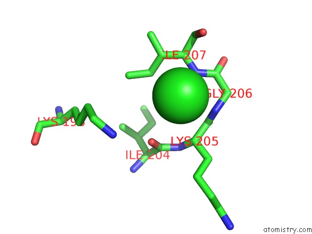
Mono view
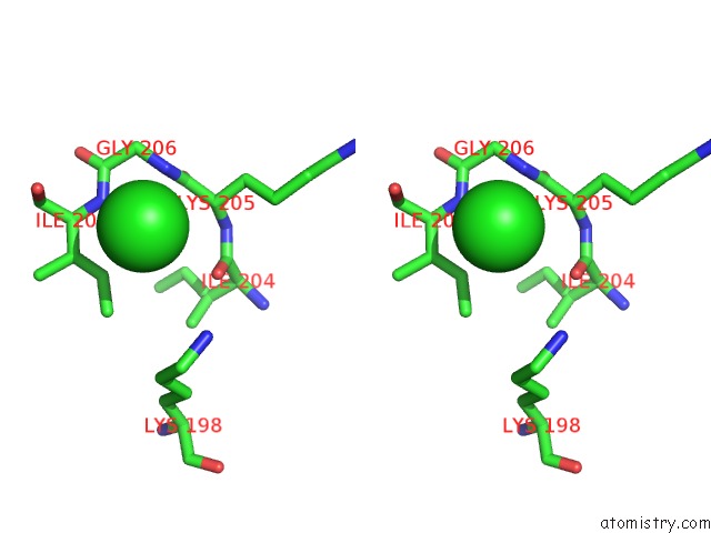
Stereo pair view
Mono view
Stereo pair view
A full contact list of Chlorine with other atoms in the Cl binding
site number 1 of 2.45 Angstrom Resolution Crystal Structure of Udp-N-Acetylglucosamine 1-Carboxyvinyltransferase From Campylobacter Jejuni. within 5.0Å range:
|
Reference:
G.Minasov,
L.Shuvalova,
I.Dubrovska,
J.Winsor,
J.Stam,
K.Kwon,
W.F.Anderson,
Center For Structural Genomics Of Infectious Diseases(Csgid).
2.45 Angstrom Resolution Crystal Structure of Udp-N-Acetylglucosamine 1-Carboxyvinyltransferase From Campylobacter Jejuni. To Be Published.
Page generated: Sat Jul 12 09:23:15 2025
Last articles
Na in 1Z1QNa in 1YYG
Na in 1YYA
Na in 1YJ9
Na in 1YX1
Na in 1YVM
Na in 1YQ2
Na in 1YPL
Na in 1YPE
Na in 1YNQ