Chlorine »
PDB 5upg-5uwx »
5uqs »
Chlorine in PDB 5uqs: Crystal Structure of Citrate Synthase From Sus Scrofa
Enzymatic activity of Crystal Structure of Citrate Synthase From Sus Scrofa
All present enzymatic activity of Crystal Structure of Citrate Synthase From Sus Scrofa:
2.3.3.1;
2.3.3.1;
Protein crystallography data
The structure of Crystal Structure of Citrate Synthase From Sus Scrofa, PDB code: 5uqs
was solved by
C.Schlachter,
M.Chruszcz,
with X-Ray Crystallography technique. A brief refinement statistics is given in the table below:
| Resolution Low / High (Å) | 34.00 / 1.60 |
| Space group | P 1 |
| Cell size a, b, c (Å), α, β, γ (°) | 58.575, 59.884, 74.761, 99.60, 98.47, 117.53 |
| R / Rfree (%) | 15.3 / 17.8 |
Chlorine Binding Sites:
The binding sites of Chlorine atom in the Crystal Structure of Citrate Synthase From Sus Scrofa
(pdb code 5uqs). This binding sites where shown within
5.0 Angstroms radius around Chlorine atom.
In total 2 binding sites of Chlorine where determined in the Crystal Structure of Citrate Synthase From Sus Scrofa, PDB code: 5uqs:
Jump to Chlorine binding site number: 1; 2;
In total 2 binding sites of Chlorine where determined in the Crystal Structure of Citrate Synthase From Sus Scrofa, PDB code: 5uqs:
Jump to Chlorine binding site number: 1; 2;
Chlorine binding site 1 out of 2 in 5uqs
Go back to
Chlorine binding site 1 out
of 2 in the Crystal Structure of Citrate Synthase From Sus Scrofa
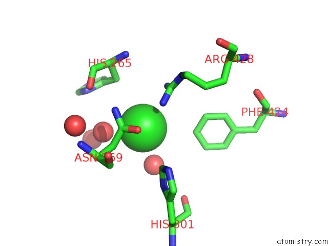
Mono view
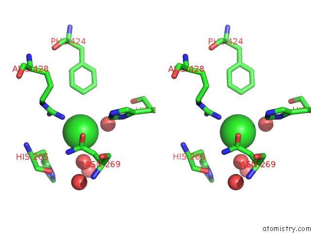
Stereo pair view
Mono view
Stereo pair view
A full contact list of Chlorine with other atoms in the Cl binding
site number 1 of Crystal Structure of Citrate Synthase From Sus Scrofa within 5.0Å range:
|
Chlorine binding site 2 out of 2 in 5uqs
Go back to
Chlorine binding site 2 out
of 2 in the Crystal Structure of Citrate Synthase From Sus Scrofa
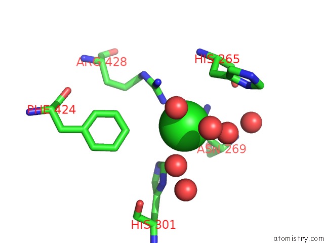
Mono view
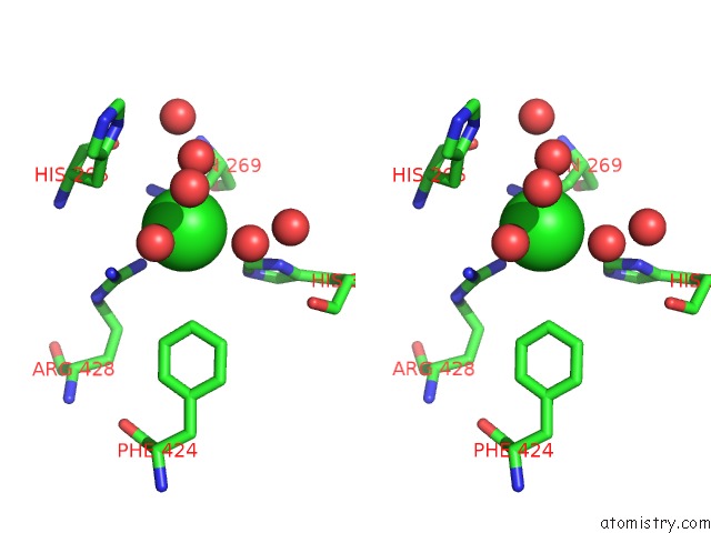
Stereo pair view
Mono view
Stereo pair view
A full contact list of Chlorine with other atoms in the Cl binding
site number 2 of Crystal Structure of Citrate Synthase From Sus Scrofa within 5.0Å range:
|
Reference:
C.Schlachter,
M.Chruszcz.
Structural and Functional Characterization of 2-Methylcitrate Synthase (Mcsa) From Aspergillus Fumigatus and Citrate Synthase (Hcs) From Homo Sapiens. To Be Published.
Page generated: Sat Jul 12 09:28:15 2025
Last articles
Na in 5YYDNa in 5Z2V
Na in 5Z1N
Na in 5YZ3
Na in 5YWV
Na in 5YU4
Na in 5YXF
Na in 5YUR
Na in 5YVE
Na in 5YU3