Chlorine »
PDB 5vvb-5w5o »
5vvi »
Chlorine in PDB 5vvi: Crystal Structure of the Ligand Binding Domain of Lysr-Type Transcriptional Regulator, Occr From Agrobacterium Tumefaciens in the Complex with Octopine
Protein crystallography data
The structure of Crystal Structure of the Ligand Binding Domain of Lysr-Type Transcriptional Regulator, Occr From Agrobacterium Tumefaciens in the Complex with Octopine, PDB code: 5vvi
was solved by
Y.Kim,
G.Chhor,
R.Jedrzejczak,
S.C.Winans,
A.Joachimiak,
Midwest Centerfor Structural Genomics (Mcsg),
with X-Ray Crystallography technique. A brief refinement statistics is given in the table below:
| Resolution Low / High (Å) | 31.84 / 2.28 |
| Space group | C 1 2 1 |
| Cell size a, b, c (Å), α, β, γ (°) | 58.417, 101.985, 112.707, 90.00, 98.14, 90.00 |
| R / Rfree (%) | 19.2 / 24.9 |
Chlorine Binding Sites:
The binding sites of Chlorine atom in the Crystal Structure of the Ligand Binding Domain of Lysr-Type Transcriptional Regulator, Occr From Agrobacterium Tumefaciens in the Complex with Octopine
(pdb code 5vvi). This binding sites where shown within
5.0 Angstroms radius around Chlorine atom.
In total only one binding site of Chlorine was determined in the Crystal Structure of the Ligand Binding Domain of Lysr-Type Transcriptional Regulator, Occr From Agrobacterium Tumefaciens in the Complex with Octopine, PDB code: 5vvi:
In total only one binding site of Chlorine was determined in the Crystal Structure of the Ligand Binding Domain of Lysr-Type Transcriptional Regulator, Occr From Agrobacterium Tumefaciens in the Complex with Octopine, PDB code: 5vvi:
Chlorine binding site 1 out of 1 in 5vvi
Go back to
Chlorine binding site 1 out
of 1 in the Crystal Structure of the Ligand Binding Domain of Lysr-Type Transcriptional Regulator, Occr From Agrobacterium Tumefaciens in the Complex with Octopine
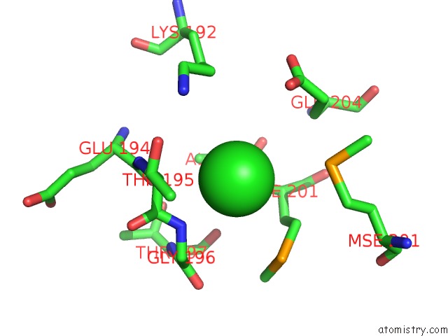
Mono view
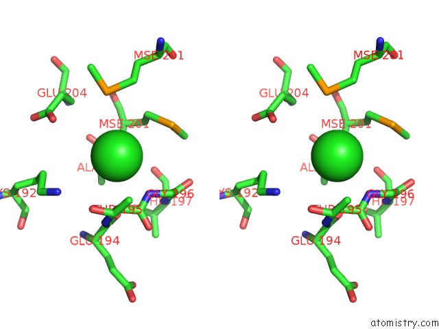
Stereo pair view
Mono view
Stereo pair view
A full contact list of Chlorine with other atoms in the Cl binding
site number 1 of Crystal Structure of the Ligand Binding Domain of Lysr-Type Transcriptional Regulator, Occr From Agrobacterium Tumefaciens in the Complex with Octopine within 5.0Å range:
|
Reference:
Y.Kim,
G.Chhor,
C.S.Tsai,
J.B.Winans,
R.Jedrzejczak,
A.Joachimiak,
S.C.Winans.
Crystal Structure of the Ligand-Binding Domain of A Lysr-Type Transcriptional Regulator: Transcriptional Activation Via A Rotary Switch. Mol. Microbiol. 2018.
ISSN: ESSN 1365-2958
PubMed: 30168204
DOI: 10.1111/MMI.14115
Page generated: Sat Jul 12 10:03:54 2025
ISSN: ESSN 1365-2958
PubMed: 30168204
DOI: 10.1111/MMI.14115
Last articles
K in 3QJ5K in 3QHD
K in 3QH8
K in 3QEK
K in 3QE9
K in 3QCR
K in 3QC5
K in 3Q9B
K in 3POX
K in 3Q8M