Chlorine »
PDB 5w5s-5web »
5w71 »
Chlorine in PDB 5w71: X-Ray Structure of Btrr From Bacillus Circulans in the Presence of the 2-Dos External Aldimine
Enzymatic activity of X-Ray Structure of Btrr From Bacillus Circulans in the Presence of the 2-Dos External Aldimine
All present enzymatic activity of X-Ray Structure of Btrr From Bacillus Circulans in the Presence of the 2-Dos External Aldimine:
2.6.1.100; 2.6.1.101;
2.6.1.100; 2.6.1.101;
Protein crystallography data
The structure of X-Ray Structure of Btrr From Bacillus Circulans in the Presence of the 2-Dos External Aldimine, PDB code: 5w71
was solved by
T.R.Zachman-Brockmeyer,
J.B.Thoden,
H.M.Holden,
with X-Ray Crystallography technique. A brief refinement statistics is given in the table below:
| Resolution Low / High (Å) | 50.00 / 2.10 |
| Space group | P 21 21 21 |
| Cell size a, b, c (Å), α, β, γ (°) | 69.400, 74.800, 180.100, 90.00, 90.00, 90.00 |
| R / Rfree (%) | 19 / 23.9 |
Chlorine Binding Sites:
The binding sites of Chlorine atom in the X-Ray Structure of Btrr From Bacillus Circulans in the Presence of the 2-Dos External Aldimine
(pdb code 5w71). This binding sites where shown within
5.0 Angstroms radius around Chlorine atom.
In total only one binding site of Chlorine was determined in the X-Ray Structure of Btrr From Bacillus Circulans in the Presence of the 2-Dos External Aldimine, PDB code: 5w71:
In total only one binding site of Chlorine was determined in the X-Ray Structure of Btrr From Bacillus Circulans in the Presence of the 2-Dos External Aldimine, PDB code: 5w71:
Chlorine binding site 1 out of 1 in 5w71
Go back to
Chlorine binding site 1 out
of 1 in the X-Ray Structure of Btrr From Bacillus Circulans in the Presence of the 2-Dos External Aldimine
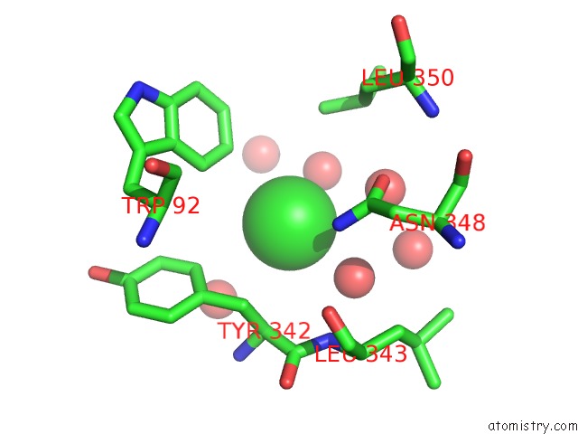
Mono view
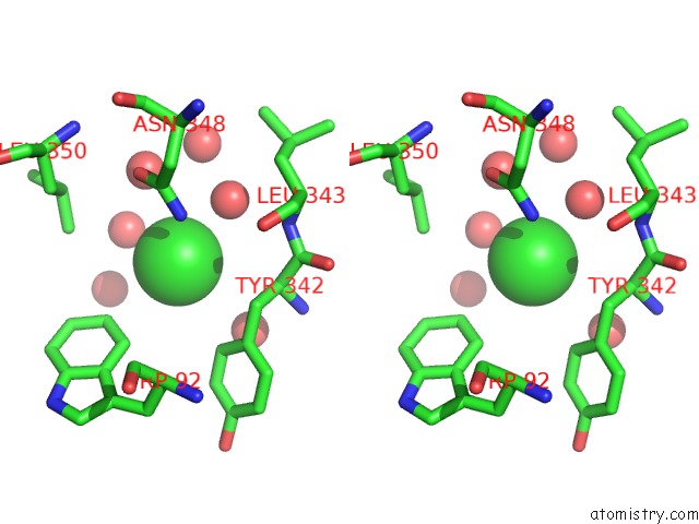
Stereo pair view
Mono view
Stereo pair view
A full contact list of Chlorine with other atoms in the Cl binding
site number 1 of X-Ray Structure of Btrr From Bacillus Circulans in the Presence of the 2-Dos External Aldimine within 5.0Å range:
|
Reference:
T.R.Zachman-Brockmeyer,
J.B.Thoden,
H.M.Holden.
The Structure of Rbmb From Streptomyces Ribosidificus, An Aminotransferase Involved in the Biosynthesis of Ribostamycin. Protein Sci. V. 26 1886 2017.
ISSN: ESSN 1469-896X
PubMed: 28685903
DOI: 10.1002/PRO.3221
Page generated: Sat Jul 12 10:10:23 2025
ISSN: ESSN 1469-896X
PubMed: 28685903
DOI: 10.1002/PRO.3221
Last articles
Mg in 3G73Mg in 3G37
Mg in 3G6Y
Mg in 3G6X
Mg in 3G6W
Mg in 3G6V
Mg in 3G6K
Mg in 3G5A
Mg in 3G5S
Mg in 3G58