Chlorine »
PDB 5xvu-5y7x »
5xyo »
Chlorine in PDB 5xyo: Structure of 6-Aminohexanoate-Oligomer Hydrolase From Arthrobacter Sp. KI72., D122G Mutant
Protein crystallography data
The structure of Structure of 6-Aminohexanoate-Oligomer Hydrolase From Arthrobacter Sp. KI72., D122G Mutant, PDB code: 5xyo
was solved by
S.Negoro,
N.Shibata,
K.Nagai,
Y.Higuchi,
with X-Ray Crystallography technique. A brief refinement statistics is given in the table below:
| Resolution Low / High (Å) | 72.23 / 2.00 |
| Space group | C 2 2 21 |
| Cell size a, b, c (Å), α, β, γ (°) | 70.485, 144.459, 128.353, 90.00, 90.00, 90.00 |
| R / Rfree (%) | 17.3 / 22.6 |
Chlorine Binding Sites:
The binding sites of Chlorine atom in the Structure of 6-Aminohexanoate-Oligomer Hydrolase From Arthrobacter Sp. KI72., D122G Mutant
(pdb code 5xyo). This binding sites where shown within
5.0 Angstroms radius around Chlorine atom.
In total 2 binding sites of Chlorine where determined in the Structure of 6-Aminohexanoate-Oligomer Hydrolase From Arthrobacter Sp. KI72., D122G Mutant, PDB code: 5xyo:
Jump to Chlorine binding site number: 1; 2;
In total 2 binding sites of Chlorine where determined in the Structure of 6-Aminohexanoate-Oligomer Hydrolase From Arthrobacter Sp. KI72., D122G Mutant, PDB code: 5xyo:
Jump to Chlorine binding site number: 1; 2;
Chlorine binding site 1 out of 2 in 5xyo
Go back to
Chlorine binding site 1 out
of 2 in the Structure of 6-Aminohexanoate-Oligomer Hydrolase From Arthrobacter Sp. KI72., D122G Mutant
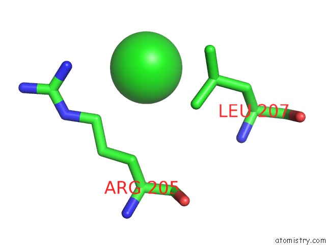
Mono view
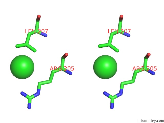
Stereo pair view
Mono view
Stereo pair view
A full contact list of Chlorine with other atoms in the Cl binding
site number 1 of Structure of 6-Aminohexanoate-Oligomer Hydrolase From Arthrobacter Sp. KI72., D122G Mutant within 5.0Å range:
|
Chlorine binding site 2 out of 2 in 5xyo
Go back to
Chlorine binding site 2 out
of 2 in the Structure of 6-Aminohexanoate-Oligomer Hydrolase From Arthrobacter Sp. KI72., D122G Mutant
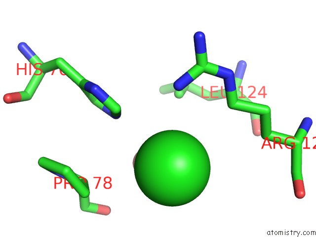
Mono view
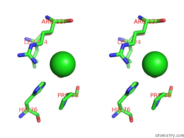
Stereo pair view
Mono view
Stereo pair view
A full contact list of Chlorine with other atoms in the Cl binding
site number 2 of Structure of 6-Aminohexanoate-Oligomer Hydrolase From Arthrobacter Sp. KI72., D122G Mutant within 5.0Å range:
|
Reference:
S.Negoro,
N.Shibata,
Y.H.Lee,
I.Takehara,
R.Kinugasa,
K.Nagai,
Y.Tanaka,
D.I.Kato,
M.Takeo,
Y.Goto,
Y.Higuchi.
Structural Basis of the Correct Subunit Assembly, Aggregation, and Intracellular Degradation of Nylon Hydrolase Sci Rep V. 8 9725 2018.
ISSN: ESSN 2045-2322
PubMed: 29950566
DOI: 10.1038/S41598-018-27860-W
Page generated: Sat Jul 12 10:48:23 2025
ISSN: ESSN 2045-2322
PubMed: 29950566
DOI: 10.1038/S41598-018-27860-W
Last articles
Mg in 4HGOMg in 4HGN
Mg in 4HGP
Mg in 4HE2
Mg in 4HEC
Mg in 4HE0
Mg in 4HEO
Mg in 4HE1
Mg in 4HCL
Mg in 4HDQ