Chlorine »
PDB 6cug-6d3i »
6czs »
Chlorine in PDB 6czs: Crystal Structure of Human Pro-Cathepsin H C26S Mutant
Enzymatic activity of Crystal Structure of Human Pro-Cathepsin H C26S Mutant
All present enzymatic activity of Crystal Structure of Human Pro-Cathepsin H C26S Mutant:
3.4.22.16;
3.4.22.16;
Protein crystallography data
The structure of Crystal Structure of Human Pro-Cathepsin H C26S Mutant, PDB code: 6czs
was solved by
X.Huang,
Y.Hao,
with X-Ray Crystallography technique. A brief refinement statistics is given in the table below:
| Resolution Low / High (Å) | 49.12 / 1.66 |
| Space group | P 31 2 1 |
| Cell size a, b, c (Å), α, β, γ (°) | 98.242, 98.242, 106.586, 90.00, 90.00, 120.00 |
| R / Rfree (%) | 18.1 / 20.9 |
Chlorine Binding Sites:
The binding sites of Chlorine atom in the Crystal Structure of Human Pro-Cathepsin H C26S Mutant
(pdb code 6czs). This binding sites where shown within
5.0 Angstroms radius around Chlorine atom.
In total 2 binding sites of Chlorine where determined in the Crystal Structure of Human Pro-Cathepsin H C26S Mutant, PDB code: 6czs:
Jump to Chlorine binding site number: 1; 2;
In total 2 binding sites of Chlorine where determined in the Crystal Structure of Human Pro-Cathepsin H C26S Mutant, PDB code: 6czs:
Jump to Chlorine binding site number: 1; 2;
Chlorine binding site 1 out of 2 in 6czs
Go back to
Chlorine binding site 1 out
of 2 in the Crystal Structure of Human Pro-Cathepsin H C26S Mutant
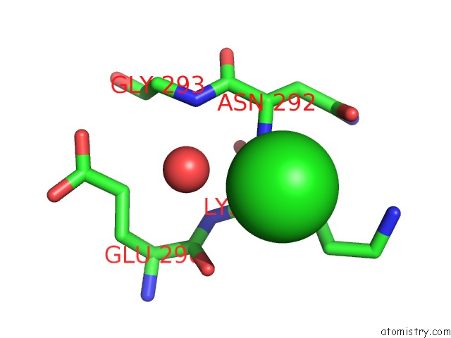
Mono view
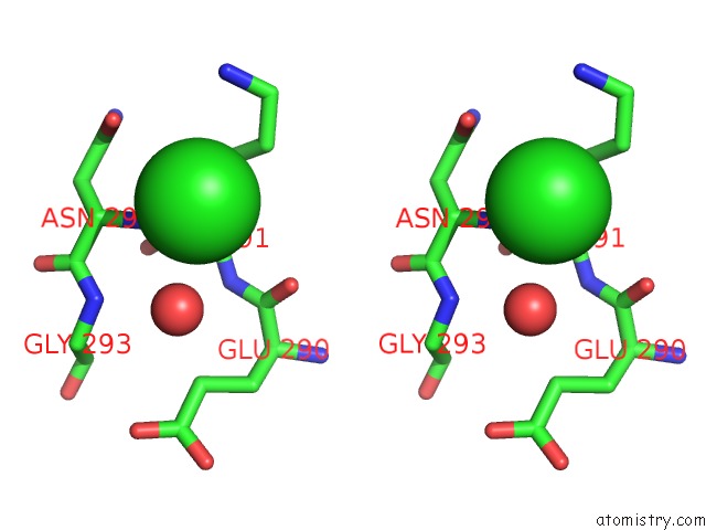
Stereo pair view
Mono view
Stereo pair view
A full contact list of Chlorine with other atoms in the Cl binding
site number 1 of Crystal Structure of Human Pro-Cathepsin H C26S Mutant within 5.0Å range:
|
Chlorine binding site 2 out of 2 in 6czs
Go back to
Chlorine binding site 2 out
of 2 in the Crystal Structure of Human Pro-Cathepsin H C26S Mutant
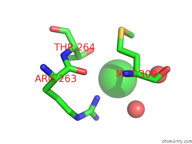
Mono view
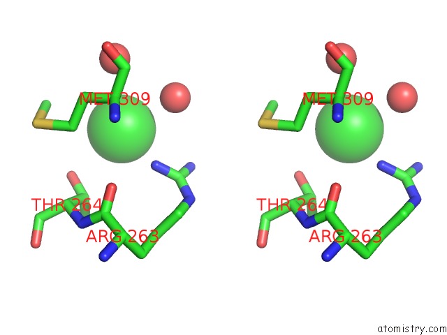
Stereo pair view
Mono view
Stereo pair view
A full contact list of Chlorine with other atoms in the Cl binding
site number 2 of Crystal Structure of Human Pro-Cathepsin H C26S Mutant within 5.0Å range:
|
Reference:
Y.Hao,
W.Purtha,
C.Cortesio,
H.Rui,
Y.Gu,
H.Chen,
E.A.Sickmier,
P.Manzanillo,
X.Huang.
Crystal Structures of Human Procathepsin H. Plos One V. 13 00374 2018.
ISSN: ESSN 1932-6203
PubMed: 30044821
DOI: 10.1371/JOURNAL.PONE.0200374
Page generated: Sat Jul 12 12:37:24 2025
ISSN: ESSN 1932-6203
PubMed: 30044821
DOI: 10.1371/JOURNAL.PONE.0200374
Last articles
Mg in 1ZSZMg in 1ZUN
Mg in 1ZUK
Mg in 1ZPD
Mg in 1ZS9
Mg in 1ZOT
Mg in 1ZSH
Mg in 1ZPI
Mg in 1ZOO
Mg in 1ZPH