Chlorine »
PDB 6d3m-6de4 »
6d4c »
Chlorine in PDB 6d4c: Crystal Structure of Candida Boidinii Formate Dehydrogenase V123G Mutant Complexed with Nad+ and Azide
Enzymatic activity of Crystal Structure of Candida Boidinii Formate Dehydrogenase V123G Mutant Complexed with Nad+ and Azide
All present enzymatic activity of Crystal Structure of Candida Boidinii Formate Dehydrogenase V123G Mutant Complexed with Nad+ and Azide:
1.17.1.9;
1.17.1.9;
Protein crystallography data
The structure of Crystal Structure of Candida Boidinii Formate Dehydrogenase V123G Mutant Complexed with Nad+ and Azide, PDB code: 6d4c
was solved by
Q.Guo,
H.Ye,
L.Gakhar,
C.M.Cheatum,
A.Kohen,
with X-Ray Crystallography technique. A brief refinement statistics is given in the table below:
| Resolution Low / High (Å) | 45.99 / 1.45 |
| Space group | P 1 21 1 |
| Cell size a, b, c (Å), α, β, γ (°) | 51.188, 115.899, 65.469, 90.00, 108.56, 90.00 |
| R / Rfree (%) | 15.9 / 18.5 |
Chlorine Binding Sites:
The binding sites of Chlorine atom in the Crystal Structure of Candida Boidinii Formate Dehydrogenase V123G Mutant Complexed with Nad+ and Azide
(pdb code 6d4c). This binding sites where shown within
5.0 Angstroms radius around Chlorine atom.
In total 2 binding sites of Chlorine where determined in the Crystal Structure of Candida Boidinii Formate Dehydrogenase V123G Mutant Complexed with Nad+ and Azide, PDB code: 6d4c:
Jump to Chlorine binding site number: 1; 2;
In total 2 binding sites of Chlorine where determined in the Crystal Structure of Candida Boidinii Formate Dehydrogenase V123G Mutant Complexed with Nad+ and Azide, PDB code: 6d4c:
Jump to Chlorine binding site number: 1; 2;
Chlorine binding site 1 out of 2 in 6d4c
Go back to
Chlorine binding site 1 out
of 2 in the Crystal Structure of Candida Boidinii Formate Dehydrogenase V123G Mutant Complexed with Nad+ and Azide
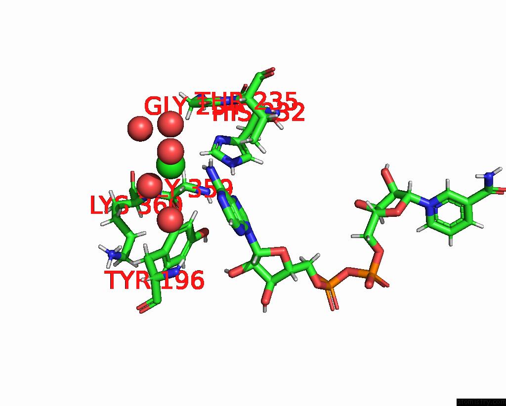
Mono view
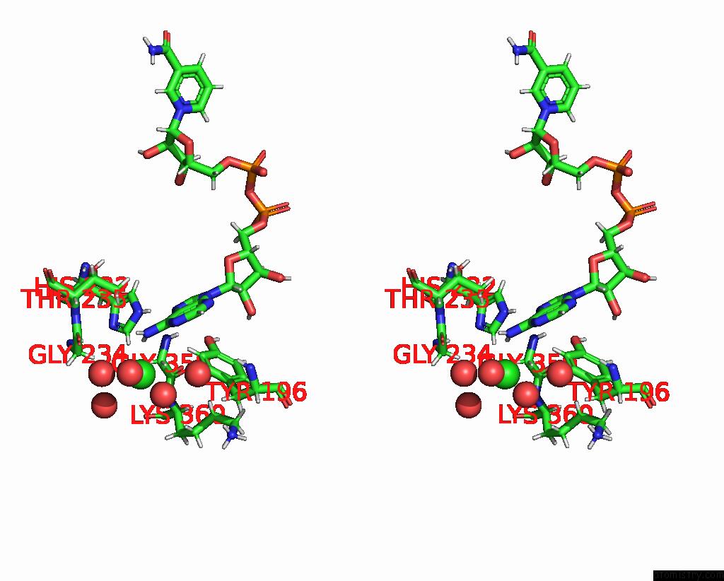
Stereo pair view
Mono view
Stereo pair view
A full contact list of Chlorine with other atoms in the Cl binding
site number 1 of Crystal Structure of Candida Boidinii Formate Dehydrogenase V123G Mutant Complexed with Nad+ and Azide within 5.0Å range:
|
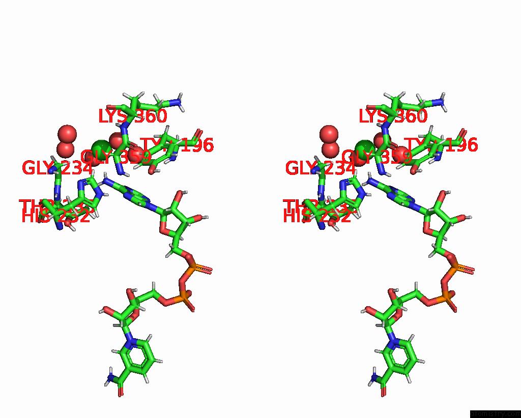
Chlorine binding site 2 out of 2 in 6d4c
Go back to
Chlorine binding site 2 out
of 2 in the Crystal Structure of Candida Boidinii Formate Dehydrogenase V123G Mutant Complexed with Nad+ and Azide
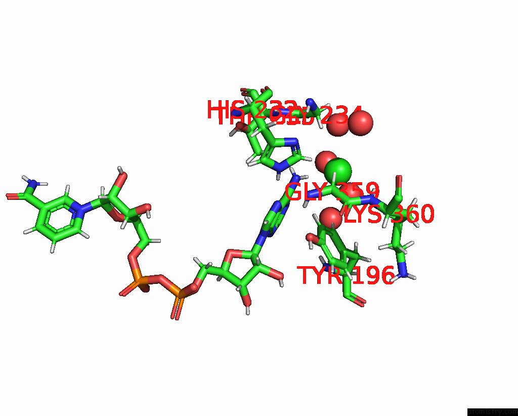
Mono view
Stereo pair view
Mono view
Stereo pair view
A full contact list of Chlorine with other atoms in the Cl binding
site number 2 of Crystal Structure of Candida Boidinii Formate Dehydrogenase V123G Mutant Complexed with Nad+ and Azide within 5.0Å range:
|
Reference:
P.L.Pagano,
Q.Guo,
C.Ranasinghe,
E.Schroeder,
K.Robben,
F.Hase,
H.Ye,
K.Wickersham,
A.Aspuru-Guzik,
D.T.Major,
L.Gakhar,
A.Kohen,
C.M.Cheatum.
Oscillatory Active-Site Motions Correlate with Kinetic Isotope Effects in Formate Dehydrogenase Acs Catalysis 2019.
ISSN: ESSN 2155-5435
DOI: 10.1021/ACSCATAL.9B03345
Page generated: Sat Jul 12 12:43:13 2025
ISSN: ESSN 2155-5435
DOI: 10.1021/ACSCATAL.9B03345
Last articles
Mg in 5Y8HMg in 5Y8B
Mg in 5Y5P
Mg in 5Y5Q
Mg in 5Y6Z
Mg in 5XYM
Mg in 5XYU
Mg in 5Y4N
Mg in 5Y4J
Mg in 5Y4I