Chlorine »
PDB 6dox-6dy2 »
6ds8 »
Chlorine in PDB 6ds8: Crystal Structure of Mhud R26S Mutant with Two Manganese Protoporphyrin IX Bound Per Active Site
Enzymatic activity of Crystal Structure of Mhud R26S Mutant with Two Manganese Protoporphyrin IX Bound Per Active Site
All present enzymatic activity of Crystal Structure of Mhud R26S Mutant with Two Manganese Protoporphyrin IX Bound Per Active Site:
1.14.14.18;
1.14.14.18;
Protein crystallography data
The structure of Crystal Structure of Mhud R26S Mutant with Two Manganese Protoporphyrin IX Bound Per Active Site, PDB code: 6ds8
was solved by
A.Chao,
C.W.Goulding,
with X-Ray Crystallography technique. A brief refinement statistics is given in the table below:
| Resolution Low / High (Å) | 50.50 / 2.40 |
| Space group | P 21 21 21 |
| Cell size a, b, c (Å), α, β, γ (°) | 39.020, 68.701, 74.490, 90.00, 90.00, 90.00 |
| R / Rfree (%) | 18.1 / 24.2 |
Other elements in 6ds8:
The structure of Crystal Structure of Mhud R26S Mutant with Two Manganese Protoporphyrin IX Bound Per Active Site also contains other interesting chemical elements:
| Manganese | (Mn) | 4 atoms |
Chlorine Binding Sites:
The binding sites of Chlorine atom in the Crystal Structure of Mhud R26S Mutant with Two Manganese Protoporphyrin IX Bound Per Active Site
(pdb code 6ds8). This binding sites where shown within
5.0 Angstroms radius around Chlorine atom.
In total 2 binding sites of Chlorine where determined in the Crystal Structure of Mhud R26S Mutant with Two Manganese Protoporphyrin IX Bound Per Active Site, PDB code: 6ds8:
Jump to Chlorine binding site number: 1; 2;
In total 2 binding sites of Chlorine where determined in the Crystal Structure of Mhud R26S Mutant with Two Manganese Protoporphyrin IX Bound Per Active Site, PDB code: 6ds8:
Jump to Chlorine binding site number: 1; 2;
Chlorine binding site 1 out of 2 in 6ds8
Go back to
Chlorine binding site 1 out
of 2 in the Crystal Structure of Mhud R26S Mutant with Two Manganese Protoporphyrin IX Bound Per Active Site

Mono view
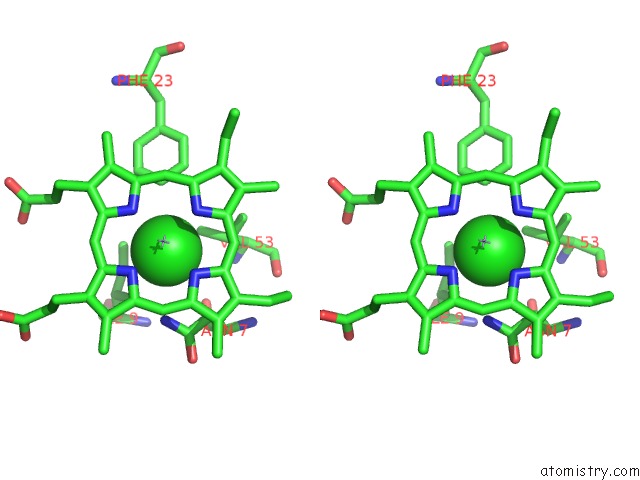
Stereo pair view
Mono view
Stereo pair view
A full contact list of Chlorine with other atoms in the Cl binding
site number 1 of Crystal Structure of Mhud R26S Mutant with Two Manganese Protoporphyrin IX Bound Per Active Site within 5.0Å range:
|
Chlorine binding site 2 out of 2 in 6ds8
Go back to
Chlorine binding site 2 out
of 2 in the Crystal Structure of Mhud R26S Mutant with Two Manganese Protoporphyrin IX Bound Per Active Site

Mono view
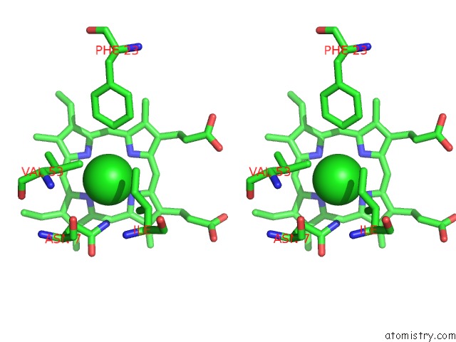
Stereo pair view
Mono view
Stereo pair view
A full contact list of Chlorine with other atoms in the Cl binding
site number 2 of Crystal Structure of Mhud R26S Mutant with Two Manganese Protoporphyrin IX Bound Per Active Site within 5.0Å range:
|
Reference:
A.Chao,
C.W.Goulding.
Crystal Structure of Mhud R26S Mutant with Two Manganese Protoporphyrin IX Bound Per Active Site To Be Published.
Page generated: Sat Jul 12 13:00:54 2025
Last articles
Mg in 1VROMg in 1VRN
Mg in 1VRG
Mg in 1VR0
Mg in 1VQM
Mg in 1VQL
Mg in 1VQK
Mg in 1VQ9
Mg in 1VQ7
Mg in 1VQ5