Chlorine »
PDB 6fs0-6g1v »
6ft8 »
Chlorine in PDB 6ft8: Crystal Structure of CLK1 in Complex with Inhibitor 8G
Enzymatic activity of Crystal Structure of CLK1 in Complex with Inhibitor 8G
All present enzymatic activity of Crystal Structure of CLK1 in Complex with Inhibitor 8G:
2.7.12.1;
2.7.12.1;
Protein crystallography data
The structure of Crystal Structure of CLK1 in Complex with Inhibitor 8G, PDB code: 6ft8
was solved by
A.Chaikuad,
A.Walter,
F.Von Delft,
C.Bountra,
C.H.Arrowsmith,
A.M.Edwards,
C.Kunick,
S.Knapp,
Structural Genomics Consortium (Sgc),
with X-Ray Crystallography technique. A brief refinement statistics is given in the table below:
| Resolution Low / High (Å) | 23.65 / 1.45 |
| Space group | C 1 2 1 |
| Cell size a, b, c (Å), α, β, γ (°) | 91.640, 64.210, 88.360, 90.00, 127.67, 90.00 |
| R / Rfree (%) | 16.4 / 19.5 |
Other elements in 6ft8:
The structure of Crystal Structure of CLK1 in Complex with Inhibitor 8G also contains other interesting chemical elements:
| Sodium | (Na) | 1 atom |
Chlorine Binding Sites:
The binding sites of Chlorine atom in the Crystal Structure of CLK1 in Complex with Inhibitor 8G
(pdb code 6ft8). This binding sites where shown within
5.0 Angstroms radius around Chlorine atom.
In total only one binding site of Chlorine was determined in the Crystal Structure of CLK1 in Complex with Inhibitor 8G, PDB code: 6ft8:
In total only one binding site of Chlorine was determined in the Crystal Structure of CLK1 in Complex with Inhibitor 8G, PDB code: 6ft8:
Chlorine binding site 1 out of 1 in 6ft8
Go back to
Chlorine binding site 1 out
of 1 in the Crystal Structure of CLK1 in Complex with Inhibitor 8G
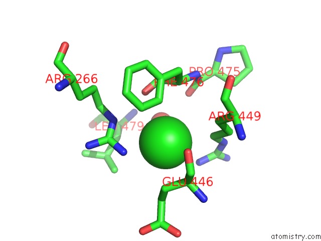
Mono view
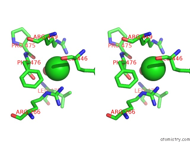
Stereo pair view
Mono view
Stereo pair view
A full contact list of Chlorine with other atoms in the Cl binding
site number 1 of Crystal Structure of CLK1 in Complex with Inhibitor 8G within 5.0Å range:
|
Reference:
A.Walter,
A.Chaikuad,
R.Helmer,
N.Loaec,
L.Preu,
I.Ott,
S.Knapp,
L.Meijer,
C.Kunick.
Molecular Structures of CDC2-Like Kinases in Complex with A New Inhibitor Chemotype. Plos One V. 13 96761 2018.
ISSN: ESSN 1932-6203
PubMed: 29723265
DOI: 10.1371/JOURNAL.PONE.0196761
Page generated: Sat Jul 12 14:13:54 2025
ISSN: ESSN 1932-6203
PubMed: 29723265
DOI: 10.1371/JOURNAL.PONE.0196761
Last articles
Mn in 8IRDMn in 8IRB
Mn in 8IRA
Mn in 8IRC
Mn in 8IR9
Mn in 8IR8
Mn in 8IR7
Mn in 8IR6
Mn in 8IR5
Mn in 8IQD