Chlorine »
PDB 6fs0-6g1v »
6fz4 »
Chlorine in PDB 6fz4: Structure of GLUK1 Ligand-Binding Domain in Complex with N-(7-Fluoro- 2,3-Dioxo-6-(Trifluoromethyl)-3,4-Dihydroquinoxalin-1(2H)-Yl)-2- Hydroxybenzamide at 1.85 A Resolution
Protein crystallography data
The structure of Structure of GLUK1 Ligand-Binding Domain in Complex with N-(7-Fluoro- 2,3-Dioxo-6-(Trifluoromethyl)-3,4-Dihydroquinoxalin-1(2H)-Yl)-2- Hydroxybenzamide at 1.85 A Resolution, PDB code: 6fz4
was solved by
J.S.Kastrup,
K.Frydenvang,
S.Mollerud,
with X-Ray Crystallography technique. A brief refinement statistics is given in the table below:
| Resolution Low / High (Å) | 37.38 / 1.85 |
| Space group | H 3 2 |
| Cell size a, b, c (Å), α, β, γ (°) | 88.867, 88.867, 157.145, 90.00, 90.00, 120.00 |
| R / Rfree (%) | 18.7 / 21.6 |
Other elements in 6fz4:
The structure of Structure of GLUK1 Ligand-Binding Domain in Complex with N-(7-Fluoro- 2,3-Dioxo-6-(Trifluoromethyl)-3,4-Dihydroquinoxalin-1(2H)-Yl)-2- Hydroxybenzamide at 1.85 A Resolution also contains other interesting chemical elements:
| Fluorine | (F) | 4 atoms |
Chlorine Binding Sites:
The binding sites of Chlorine atom in the Structure of GLUK1 Ligand-Binding Domain in Complex with N-(7-Fluoro- 2,3-Dioxo-6-(Trifluoromethyl)-3,4-Dihydroquinoxalin-1(2H)-Yl)-2- Hydroxybenzamide at 1.85 A Resolution
(pdb code 6fz4). This binding sites where shown within
5.0 Angstroms radius around Chlorine atom.
In total only one binding site of Chlorine was determined in the Structure of GLUK1 Ligand-Binding Domain in Complex with N-(7-Fluoro- 2,3-Dioxo-6-(Trifluoromethyl)-3,4-Dihydroquinoxalin-1(2H)-Yl)-2- Hydroxybenzamide at 1.85 A Resolution, PDB code: 6fz4:
In total only one binding site of Chlorine was determined in the Structure of GLUK1 Ligand-Binding Domain in Complex with N-(7-Fluoro- 2,3-Dioxo-6-(Trifluoromethyl)-3,4-Dihydroquinoxalin-1(2H)-Yl)-2- Hydroxybenzamide at 1.85 A Resolution, PDB code: 6fz4:
Chlorine binding site 1 out of 1 in 6fz4
Go back to
Chlorine binding site 1 out
of 1 in the Structure of GLUK1 Ligand-Binding Domain in Complex with N-(7-Fluoro- 2,3-Dioxo-6-(Trifluoromethyl)-3,4-Dihydroquinoxalin-1(2H)-Yl)-2- Hydroxybenzamide at 1.85 A Resolution
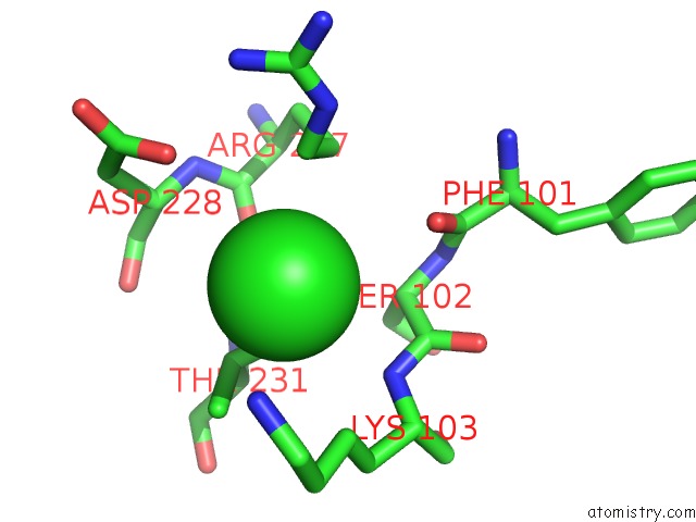
Mono view
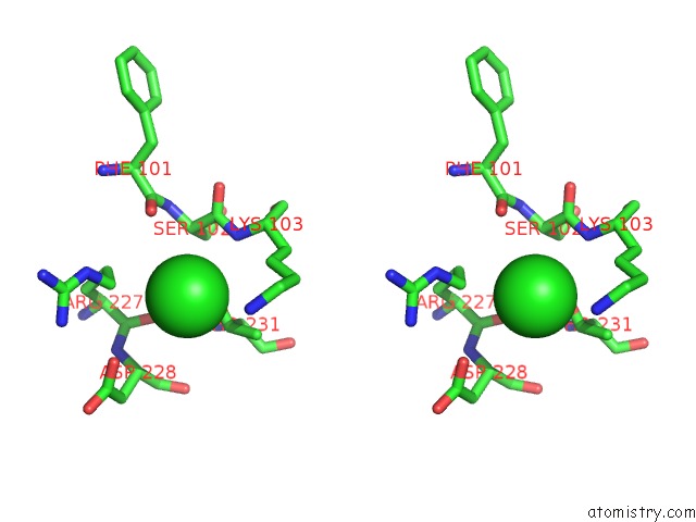
Stereo pair view
Mono view
Stereo pair view
A full contact list of Chlorine with other atoms in the Cl binding
site number 1 of Structure of GLUK1 Ligand-Binding Domain in Complex with N-(7-Fluoro- 2,3-Dioxo-6-(Trifluoromethyl)-3,4-Dihydroquinoxalin-1(2H)-Yl)-2- Hydroxybenzamide at 1.85 A Resolution within 5.0Å range:
|
Reference:
J.Pallesen,
S.Mollerud,
K.Frydenvang,
D.S.Pickering,
J.Bornholdt,
B.Nielsen,
D.Pasini,
L.Han,
L.Marconi,
J.S.Kastrup,
T.N.Johansen.
N1-Substituted Quinoxaline-2,3-Diones As Kainate Receptor Antagonists: X-Ray Crystallography, Structure-Affinity Relationships, and in Vitro Pharmacology. Acs Chem Neurosci V. 10 1841 2019.
ISSN: ESSN 1948-7193
PubMed: 30620174
DOI: 10.1021/ACSCHEMNEURO.8B00726
Page generated: Sat Jul 12 14:16:38 2025
ISSN: ESSN 1948-7193
PubMed: 30620174
DOI: 10.1021/ACSCHEMNEURO.8B00726
Last articles
Na in 4XONNa in 4XOJ
Na in 4XNX
Na in 4XN8
Na in 4XND
Na in 4XNU
Na in 4XNK
Na in 4XNB
Na in 4XNF
Na in 4XNA