Chlorine »
PDB 6gpu-6gxj »
6gqc »
Chlorine in PDB 6gqc: Crystal Structure of the PSMALPHA3 Peptide Mutant G16A Forming Cross- Alpha Amyloid-Like Fibril
Protein crystallography data
The structure of Crystal Structure of the PSMALPHA3 Peptide Mutant G16A Forming Cross- Alpha Amyloid-Like Fibril, PDB code: 6gqc
was solved by
M.Landau,
E.Tayeb-Fligelman,
with X-Ray Crystallography technique. A brief refinement statistics is given in the table below:
| Resolution Low / High (Å) | 14.67 / 1.40 |
| Space group | C 1 2 1 |
| Cell size a, b, c (Å), α, β, γ (°) | 53.270, 13.300, 25.800, 90.00, 111.91, 90.00 |
| R / Rfree (%) | 15.1 / 19.3 |
Other elements in 6gqc:
The structure of Crystal Structure of the PSMALPHA3 Peptide Mutant G16A Forming Cross- Alpha Amyloid-Like Fibril also contains other interesting chemical elements:
| Sodium | (Na) | 2 atoms |
Chlorine Binding Sites:
The binding sites of Chlorine atom in the Crystal Structure of the PSMALPHA3 Peptide Mutant G16A Forming Cross- Alpha Amyloid-Like Fibril
(pdb code 6gqc). This binding sites where shown within
5.0 Angstroms radius around Chlorine atom.
In total only one binding site of Chlorine was determined in the Crystal Structure of the PSMALPHA3 Peptide Mutant G16A Forming Cross- Alpha Amyloid-Like Fibril, PDB code: 6gqc:
In total only one binding site of Chlorine was determined in the Crystal Structure of the PSMALPHA3 Peptide Mutant G16A Forming Cross- Alpha Amyloid-Like Fibril, PDB code: 6gqc:
Chlorine binding site 1 out of 1 in 6gqc
Go back to
Chlorine binding site 1 out
of 1 in the Crystal Structure of the PSMALPHA3 Peptide Mutant G16A Forming Cross- Alpha Amyloid-Like Fibril
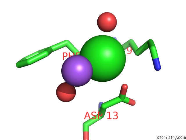
Mono view
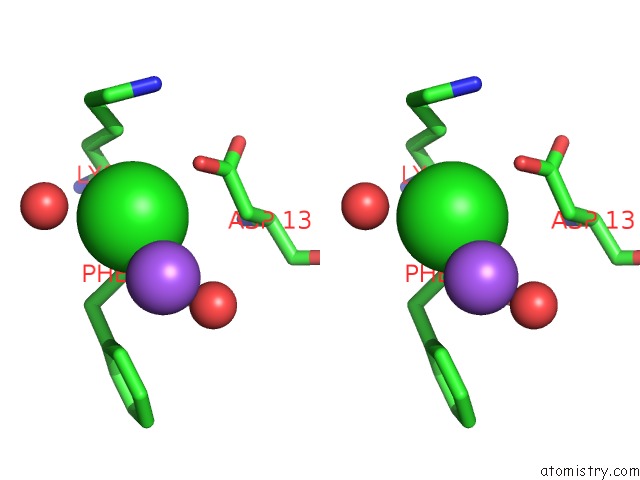
Stereo pair view
Mono view
Stereo pair view
A full contact list of Chlorine with other atoms in the Cl binding
site number 1 of Crystal Structure of the PSMALPHA3 Peptide Mutant G16A Forming Cross- Alpha Amyloid-Like Fibril within 5.0Å range:
|
Reference:
E.Tayeb-Fligelman,
M.Landau.
Structure-Fibrillation-Function Relationship of the Cytotoxic PSMALPHA3 To Be Published.
Page generated: Sat Jul 12 14:40:34 2025
Last articles
Mg in 9JMAMg in 9JKP
Mg in 9JJW
Mg in 9JJV
Mg in 9JI2
Mg in 9JFJ
Mg in 9J89
Mg in 9JC1
Mg in 9JAO
Mg in 9JA1