Chlorine »
PDB 6gxk-6h4j »
6gy5 »
Chlorine in PDB 6gy5: Crystal Structure of the Kelch Domain of Human KLHL20 in Complex with DAPK1 Peptide
Protein crystallography data
The structure of Crystal Structure of the Kelch Domain of Human KLHL20 in Complex with DAPK1 Peptide, PDB code: 6gy5
was solved by
Z.Chen,
V.Hozjan,
C.Strain-Damerell,
E.Williams,
D.Wang,
C.D.O.Cooper,
C.E.Sanvitale,
M.Fairhead,
E.P.Carpenter,
A.C.W.Pike,
T.Krojer,
V.Srikannathasan,
F.Sorrell,
C.Johansson,
S.Mathea,
N.Burgess-Brown,
F.Von Delft,
C.H.Arrowsmith,
A.M.Edwards,
C.Bountra,
A.N.Bullock,
with X-Ray Crystallography technique. A brief refinement statistics is given in the table below:
| Resolution Low / High (Å) | 29.65 / 1.09 |
| Space group | P 21 21 21 |
| Cell size a, b, c (Å), α, β, γ (°) | 40.459, 47.361, 151.933, 90.00, 90.00, 90.00 |
| R / Rfree (%) | 15.3 / 17.2 |
Other elements in 6gy5:
The structure of Crystal Structure of the Kelch Domain of Human KLHL20 in Complex with DAPK1 Peptide also contains other interesting chemical elements:
| Sodium | (Na) | 14 atoms |
Chlorine Binding Sites:
The binding sites of Chlorine atom in the Crystal Structure of the Kelch Domain of Human KLHL20 in Complex with DAPK1 Peptide
(pdb code 6gy5). This binding sites where shown within
5.0 Angstroms radius around Chlorine atom.
In total only one binding site of Chlorine was determined in the Crystal Structure of the Kelch Domain of Human KLHL20 in Complex with DAPK1 Peptide, PDB code: 6gy5:
In total only one binding site of Chlorine was determined in the Crystal Structure of the Kelch Domain of Human KLHL20 in Complex with DAPK1 Peptide, PDB code: 6gy5:
Chlorine binding site 1 out of 1 in 6gy5
Go back to
Chlorine binding site 1 out
of 1 in the Crystal Structure of the Kelch Domain of Human KLHL20 in Complex with DAPK1 Peptide
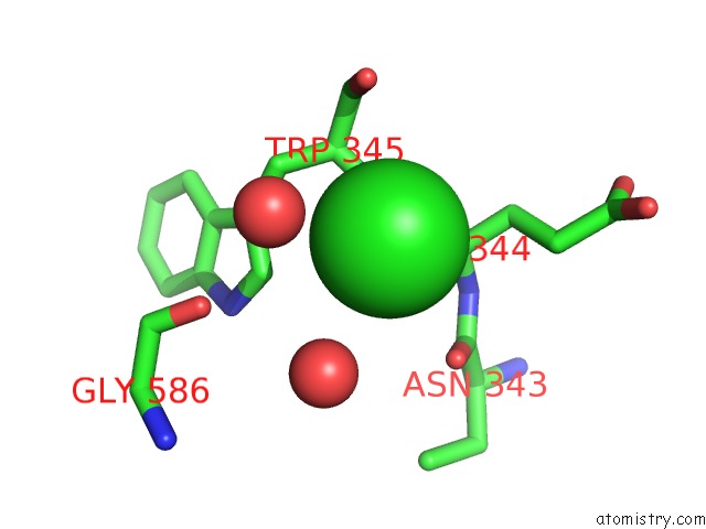
Mono view
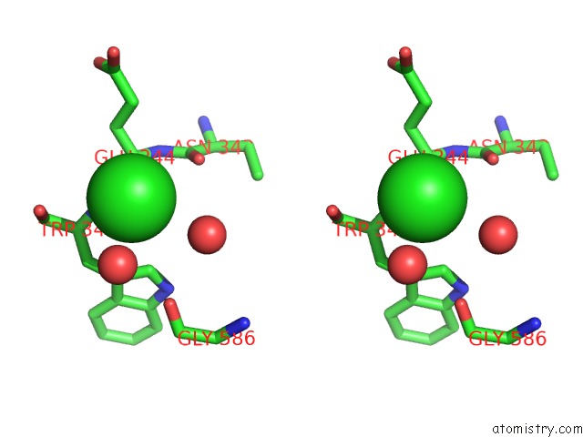
Stereo pair view
Mono view
Stereo pair view
A full contact list of Chlorine with other atoms in the Cl binding
site number 1 of Crystal Structure of the Kelch Domain of Human KLHL20 in Complex with DAPK1 Peptide within 5.0Å range:
|
Reference:
Z.Chen,
S.Picaud,
P.Filippakopoulos,
V.D'angiolella,
A.N.Bullock.
Structural Basis For Recruitment of DAPK1 to the KLHL20 E3 Ligase. Structure V. 27 1395 2019.
ISSN: ISSN 0969-2126
PubMed: 31279627
DOI: 10.1016/J.STR.2019.06.005
Page generated: Sat Jul 12 14:47:38 2025
ISSN: ISSN 0969-2126
PubMed: 31279627
DOI: 10.1016/J.STR.2019.06.005
Last articles
Mn in 4LN7Mn in 4LFI
Mn in 4LIL
Mn in 4LAC
Mn in 4L8Q
Mn in 4L78
Mn in 4KZ2
Mn in 4KPY
Mn in 4KQN
Mn in 4KQB