Chlorine »
PDB 6kkt-6kx5 »
6kol »
Chlorine in PDB 6kol: Crystal Structure of Auracyanin From Photosynthetic Bacterium Roseiflexus Castenholzii
Protein crystallography data
The structure of Crystal Structure of Auracyanin From Photosynthetic Bacterium Roseiflexus Castenholzii, PDB code: 6kol
was solved by
C.Wang,
C.Y.Zhang,
Z.Z.Min,
Y.Y.Xin,
X.L.Xu,
with X-Ray Crystallography technique. A brief refinement statistics is given in the table below:
| Resolution Low / High (Å) | 30.89 / 2.21 |
| Space group | P 2 21 21 |
| Cell size a, b, c (Å), α, β, γ (°) | 30.890, 57.912, 65.347, 90.00, 90.00, 90.00 |
| R / Rfree (%) | 18 / 23.8 |
Other elements in 6kol:
The structure of Crystal Structure of Auracyanin From Photosynthetic Bacterium Roseiflexus Castenholzii also contains other interesting chemical elements:
| Copper | (Cu) | 1 atom |
Chlorine Binding Sites:
The binding sites of Chlorine atom in the Crystal Structure of Auracyanin From Photosynthetic Bacterium Roseiflexus Castenholzii
(pdb code 6kol). This binding sites where shown within
5.0 Angstroms radius around Chlorine atom.
In total 2 binding sites of Chlorine where determined in the Crystal Structure of Auracyanin From Photosynthetic Bacterium Roseiflexus Castenholzii, PDB code: 6kol:
Jump to Chlorine binding site number: 1; 2;
In total 2 binding sites of Chlorine where determined in the Crystal Structure of Auracyanin From Photosynthetic Bacterium Roseiflexus Castenholzii, PDB code: 6kol:
Jump to Chlorine binding site number: 1; 2;
Chlorine binding site 1 out of 2 in 6kol
Go back to
Chlorine binding site 1 out
of 2 in the Crystal Structure of Auracyanin From Photosynthetic Bacterium Roseiflexus Castenholzii
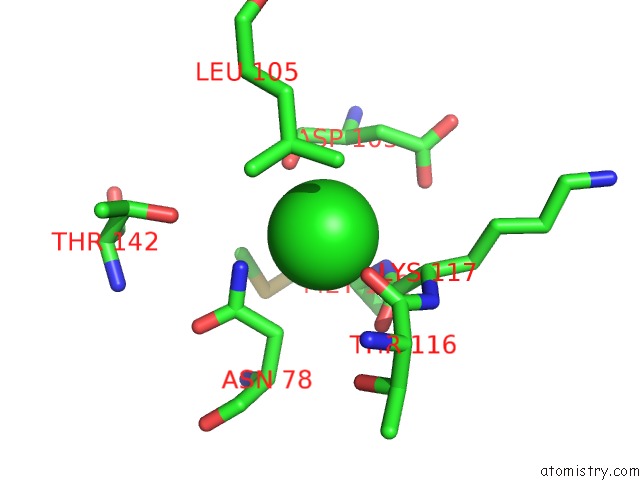
Mono view
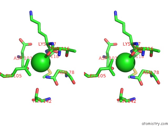
Stereo pair view
Mono view
Stereo pair view
A full contact list of Chlorine with other atoms in the Cl binding
site number 1 of Crystal Structure of Auracyanin From Photosynthetic Bacterium Roseiflexus Castenholzii within 5.0Å range:
|
Chlorine binding site 2 out of 2 in 6kol
Go back to
Chlorine binding site 2 out
of 2 in the Crystal Structure of Auracyanin From Photosynthetic Bacterium Roseiflexus Castenholzii
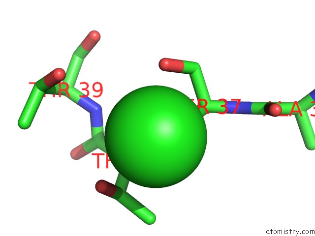
Mono view
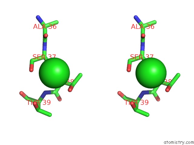
Stereo pair view
Mono view
Stereo pair view
A full contact list of Chlorine with other atoms in the Cl binding
site number 2 of Crystal Structure of Auracyanin From Photosynthetic Bacterium Roseiflexus Castenholzii within 5.0Å range:
|
Reference:
C.Wang,
Y.Y.Xin,
Z.Z.Min,
J.Qi,
C.Y.Zhang,
X.L.Xu.
Structural Basis Underlying the Electron Transfer Features of A Blue Copper Protein Auracyanin From the Photosynthetic Bacterium Roseiflexus Castenholzii. Photosyn. Res. 2020.
ISSN: ISSN 1573-5079
PubMed: 31933173
DOI: 10.1007/S11120-020-00709-Y
Page generated: Sat Jul 12 16:19:44 2025
ISSN: ISSN 1573-5079
PubMed: 31933173
DOI: 10.1007/S11120-020-00709-Y
Last articles
Mg in 2ZUVMg in 2ZUT
Mg in 2ZVJ
Mg in 2ZUW
Mg in 2ZUS
Mg in 2ZUU
Mg in 2ZTV
Mg in 2ZUE
Mg in 2ZTU
Mg in 2ZTW