Chlorine »
PDB 6kkt-6kx5 »
6kqc »
Chlorine in PDB 6kqc: Crystal Structure of E136F Mutant of Xanthine-Guanine Phosphoribosyltransferase From Yersinia Pestis
Enzymatic activity of Crystal Structure of E136F Mutant of Xanthine-Guanine Phosphoribosyltransferase From Yersinia Pestis
All present enzymatic activity of Crystal Structure of E136F Mutant of Xanthine-Guanine Phosphoribosyltransferase From Yersinia Pestis:
2.4.2.22;
2.4.2.22;
Protein crystallography data
The structure of Crystal Structure of E136F Mutant of Xanthine-Guanine Phosphoribosyltransferase From Yersinia Pestis, PDB code: 6kqc
was solved by
S.Lankipalli,
U.A.Ramagopal,
with X-Ray Crystallography technique. A brief refinement statistics is given in the table below:
| Resolution Low / High (Å) | 37.86 / 1.70 |
| Space group | P 21 21 2 |
| Cell size a, b, c (Å), α, β, γ (°) | 56.380, 96.650, 51.029, 90.00, 90.00, 90.00 |
| R / Rfree (%) | 18 / 21.2 |
Chlorine Binding Sites:
The binding sites of Chlorine atom in the Crystal Structure of E136F Mutant of Xanthine-Guanine Phosphoribosyltransferase From Yersinia Pestis
(pdb code 6kqc). This binding sites where shown within
5.0 Angstroms radius around Chlorine atom.
In total 3 binding sites of Chlorine where determined in the Crystal Structure of E136F Mutant of Xanthine-Guanine Phosphoribosyltransferase From Yersinia Pestis, PDB code: 6kqc:
Jump to Chlorine binding site number: 1; 2; 3;
In total 3 binding sites of Chlorine where determined in the Crystal Structure of E136F Mutant of Xanthine-Guanine Phosphoribosyltransferase From Yersinia Pestis, PDB code: 6kqc:
Jump to Chlorine binding site number: 1; 2; 3;
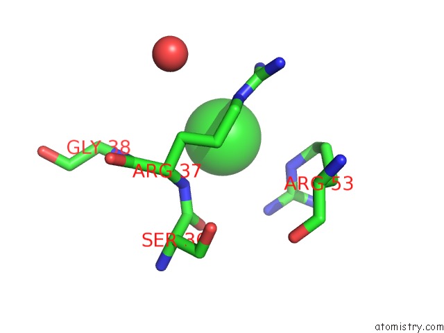
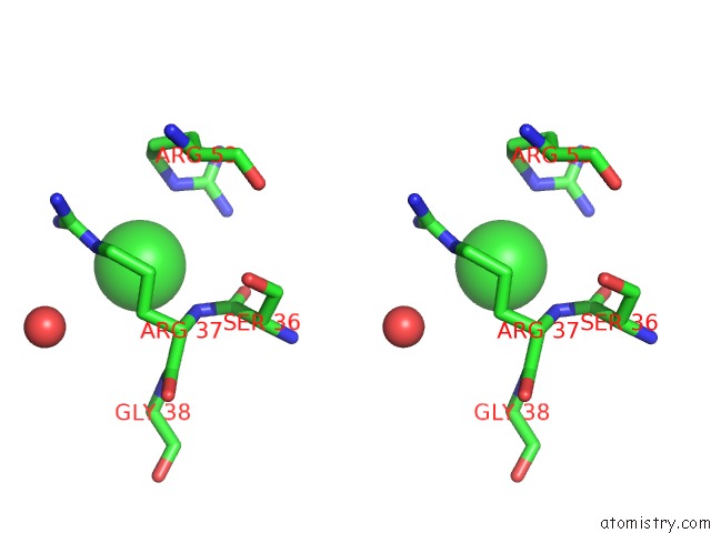
Chlorine binding site 1 out of 3 in 6kqc
Go back to
Chlorine binding site 1 out
of 3 in the Crystal Structure of E136F Mutant of Xanthine-Guanine Phosphoribosyltransferase From Yersinia Pestis
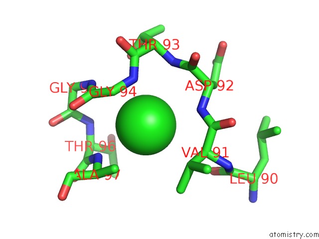
Mono view
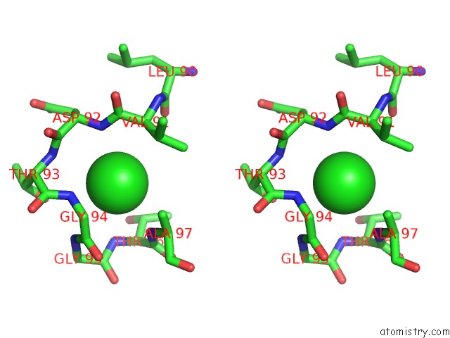
Stereo pair view
Mono view
Stereo pair view
A full contact list of Chlorine with other atoms in the Cl binding
site number 1 of Crystal Structure of E136F Mutant of Xanthine-Guanine Phosphoribosyltransferase From Yersinia Pestis within 5.0Å range:
|
Chlorine binding site 2 out of 3 in 6kqc
Go back to
Chlorine binding site 2 out
of 3 in the Crystal Structure of E136F Mutant of Xanthine-Guanine Phosphoribosyltransferase From Yersinia Pestis
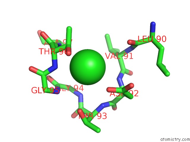
Mono view
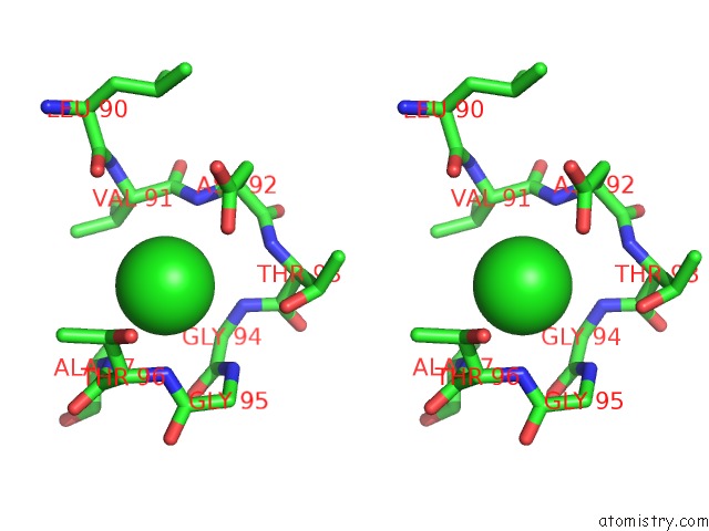
Stereo pair view
Mono view
Stereo pair view
A full contact list of Chlorine with other atoms in the Cl binding
site number 2 of Crystal Structure of E136F Mutant of Xanthine-Guanine Phosphoribosyltransferase From Yersinia Pestis within 5.0Å range:
|
Chlorine binding site 3 out of 3 in 6kqc
Go back to
Chlorine binding site 3 out
of 3 in the Crystal Structure of E136F Mutant of Xanthine-Guanine Phosphoribosyltransferase From Yersinia Pestis
Mono view
Stereo pair view
Mono view
Stereo pair view
A full contact list of Chlorine with other atoms in the Cl binding
site number 3 of Crystal Structure of E136F Mutant of Xanthine-Guanine Phosphoribosyltransferase From Yersinia Pestis within 5.0Å range:
|
Reference:
S.Lankipalli,
U.A.Ramagopal.
Crystal Structure of E136F Mutant of Xanthine-Guanine Phosphoribosyltransferase From Yersinia Pestis To Be Published.
Page generated: Sat Jul 12 16:20:46 2025
Last articles
Mg in 2Z4WMg in 2Z4T
Mg in 2Z4V
Mg in 2Z49
Mg in 2Z48
Mg in 2Z4R
Mg in 2Z4S
Mg in 2YVX
Mg in 2Z2P
Mg in 2Z41