Chlorine »
PDB 6leh-6lw2 »
6lsn »
Chlorine in PDB 6lsn: Crystal Structure of Tubulin-Inhibitor Complex
Protein crystallography data
The structure of Crystal Structure of Tubulin-Inhibitor Complex, PDB code: 6lsn
was solved by
L.Gang,
Y.X.Wang,
J.J.Cheng,
with X-Ray Crystallography technique. A brief refinement statistics is given in the table below:
| Resolution Low / High (Å) | 33.17 / 2.45 |
| Space group | P 21 21 21 |
| Cell size a, b, c (Å), α, β, γ (°) | 105.296, 158.458, 181.959, 90, 90, 90 |
| R / Rfree (%) | 17.7 / 22 |
Other elements in 6lsn:
The structure of Crystal Structure of Tubulin-Inhibitor Complex also contains other interesting chemical elements:
| Calcium | (Ca) | 4 atoms |
| Magnesium | (Mg) | 6 atoms |
Chlorine Binding Sites:
The binding sites of Chlorine atom in the Crystal Structure of Tubulin-Inhibitor Complex
(pdb code 6lsn). This binding sites where shown within
5.0 Angstroms radius around Chlorine atom.
In total only one binding site of Chlorine was determined in the Crystal Structure of Tubulin-Inhibitor Complex, PDB code: 6lsn:
In total only one binding site of Chlorine was determined in the Crystal Structure of Tubulin-Inhibitor Complex, PDB code: 6lsn:
Chlorine binding site 1 out of 1 in 6lsn
Go back to
Chlorine binding site 1 out
of 1 in the Crystal Structure of Tubulin-Inhibitor Complex
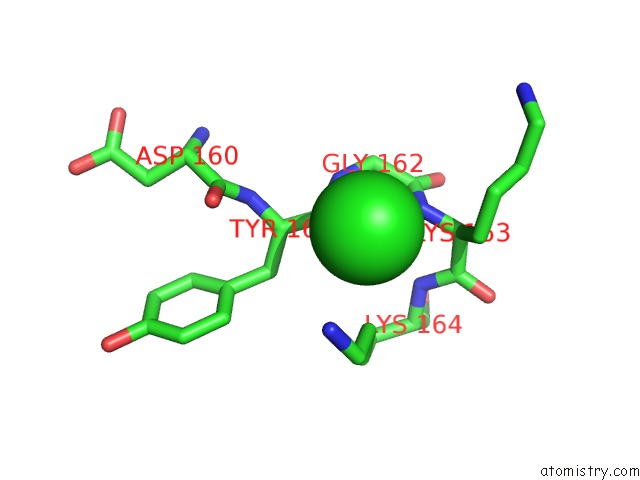
Mono view
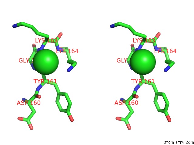
Stereo pair view
Mono view
Stereo pair view
A full contact list of Chlorine with other atoms in the Cl binding
site number 1 of Crystal Structure of Tubulin-Inhibitor Complex within 5.0Å range:
|
Reference:
L.Gang,
Y.X.Wang,
J.J.Chen.
Design, Synthesis, and Bioevaluation of Pyrazolo[1,5-A]Pyrimidine Derivatives As Tubulin Polymerization Inhibitors Targeting the Colchicine Binding Site with Potent Anticancer Activities To Be Published.
Page generated: Sat Jul 12 16:36:23 2025
Last articles
Mg in 7VUJMg in 7VUI
Mg in 7VUF
Mg in 7VSR
Mg in 7VTX
Mg in 7VTI
Mg in 7VTW
Mg in 7VTV
Mg in 7VSD
Mg in 7VSL