Chlorine »
PDB 6lx4-6m83 »
6m0f »
Chlorine in PDB 6m0f: X-Ray Structure of Drosophila Dopamine Transporter with Subsiteb Mutations (D121G/S426M) in Substrate-Free Form
Protein crystallography data
The structure of X-Ray Structure of Drosophila Dopamine Transporter with Subsiteb Mutations (D121G/S426M) in Substrate-Free Form, PDB code: 6m0f
was solved by
P.Shabareesh,
A.K.Mallela,
D.Joseph,
A.Penmatsa,
with X-Ray Crystallography technique. A brief refinement statistics is given in the table below:
| Resolution Low / High (Å) | 48.99 / 3.30 |
| Space group | P 21 21 21 |
| Cell size a, b, c (Å), α, β, γ (°) | 97.978, 133.472, 162.088, 90, 90, 90 |
| R / Rfree (%) | 24.1 / 27.7 |
Other elements in 6m0f:
The structure of X-Ray Structure of Drosophila Dopamine Transporter with Subsiteb Mutations (D121G/S426M) in Substrate-Free Form also contains other interesting chemical elements:
| Sodium | (Na) | 2 atoms |
Chlorine Binding Sites:
The binding sites of Chlorine atom in the X-Ray Structure of Drosophila Dopamine Transporter with Subsiteb Mutations (D121G/S426M) in Substrate-Free Form
(pdb code 6m0f). This binding sites where shown within
5.0 Angstroms radius around Chlorine atom.
In total only one binding site of Chlorine was determined in the X-Ray Structure of Drosophila Dopamine Transporter with Subsiteb Mutations (D121G/S426M) in Substrate-Free Form, PDB code: 6m0f:
In total only one binding site of Chlorine was determined in the X-Ray Structure of Drosophila Dopamine Transporter with Subsiteb Mutations (D121G/S426M) in Substrate-Free Form, PDB code: 6m0f:
Chlorine binding site 1 out of 1 in 6m0f
Go back to
Chlorine binding site 1 out
of 1 in the X-Ray Structure of Drosophila Dopamine Transporter with Subsiteb Mutations (D121G/S426M) in Substrate-Free Form
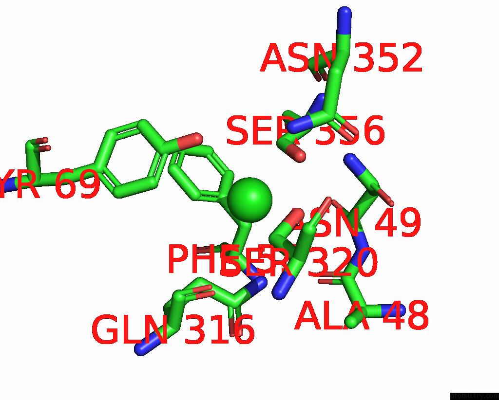
Mono view
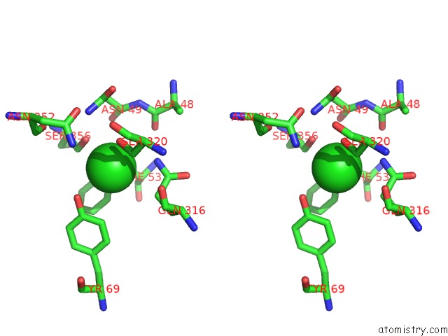
Stereo pair view
Mono view
Stereo pair view
A full contact list of Chlorine with other atoms in the Cl binding
site number 1 of X-Ray Structure of Drosophila Dopamine Transporter with Subsiteb Mutations (D121G/S426M) in Substrate-Free Form within 5.0Å range:
|
Reference:
P.Shabareesh,
A.K.Mallela,
D.Joseph.
Structural Basis of Norepinephrine Recognition and Transport Inhibition in Neurotransmitter Transporters To Be Published.
Page generated: Sat Jul 12 16:38:49 2025
Last articles
Na in 9QGBNa in 9NSM
Na in 9QGE
Na in 9P6B
Na in 9PL2
Na in 9P0F
Na in 9P4Z
Na in 9OW2
Na in 9OS1
Na in 9ORZ