Chlorine »
PDB 6m8q-6mjh »
6may »
Chlorine in PDB 6may: Crystal Structure of N-Myristoyl Transferase (Nmt) G386E Mutant From Plasmodium Vivax
Enzymatic activity of Crystal Structure of N-Myristoyl Transferase (Nmt) G386E Mutant From Plasmodium Vivax
All present enzymatic activity of Crystal Structure of N-Myristoyl Transferase (Nmt) G386E Mutant From Plasmodium Vivax:
2.3.1.97;
2.3.1.97;
Protein crystallography data
The structure of Crystal Structure of N-Myristoyl Transferase (Nmt) G386E Mutant From Plasmodium Vivax, PDB code: 6may
was solved by
Seattle Structural Genomics Center For Infectious Disease (Ssgcid),
with X-Ray Crystallography technique. A brief refinement statistics is given in the table below:
| Resolution Low / High (Å) | 50.00 / 2.05 |
| Space group | P 21 21 21 |
| Cell size a, b, c (Å), α, β, γ (°) | 57.810, 120.810, 179.060, 90.00, 90.00, 90.00 |
| R / Rfree (%) | 15.7 / 21.9 |
Chlorine Binding Sites:
The binding sites of Chlorine atom in the Crystal Structure of N-Myristoyl Transferase (Nmt) G386E Mutant From Plasmodium Vivax
(pdb code 6may). This binding sites where shown within
5.0 Angstroms radius around Chlorine atom.
In total 3 binding sites of Chlorine where determined in the Crystal Structure of N-Myristoyl Transferase (Nmt) G386E Mutant From Plasmodium Vivax, PDB code: 6may:
Jump to Chlorine binding site number: 1; 2; 3;
In total 3 binding sites of Chlorine where determined in the Crystal Structure of N-Myristoyl Transferase (Nmt) G386E Mutant From Plasmodium Vivax, PDB code: 6may:
Jump to Chlorine binding site number: 1; 2; 3;
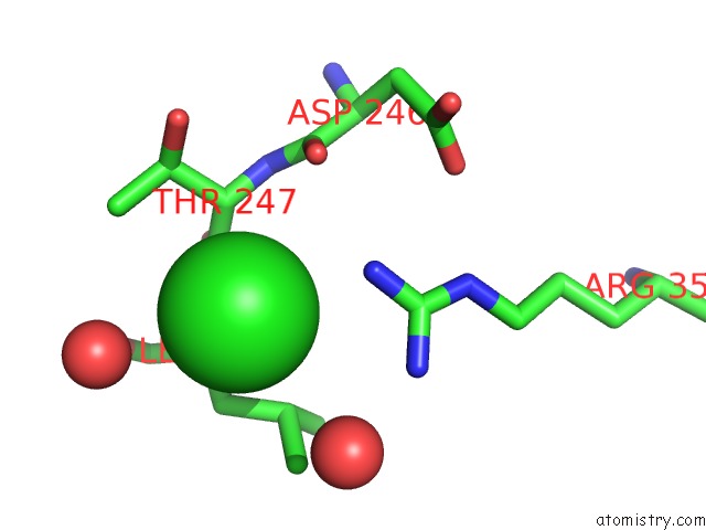
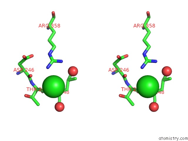
Chlorine binding site 1 out of 3 in 6may
Go back to
Chlorine binding site 1 out
of 3 in the Crystal Structure of N-Myristoyl Transferase (Nmt) G386E Mutant From Plasmodium Vivax
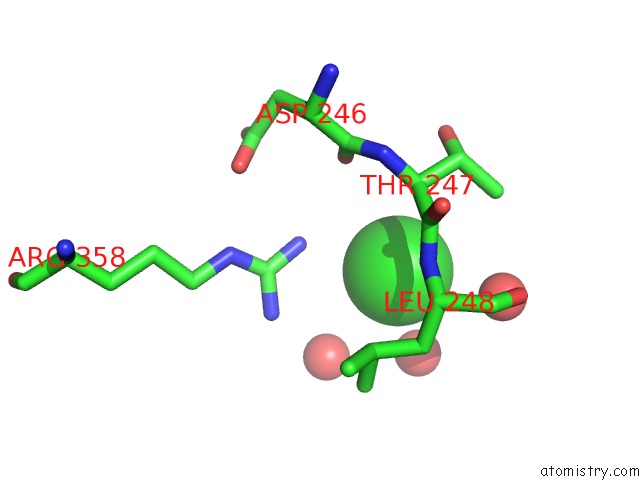
Mono view
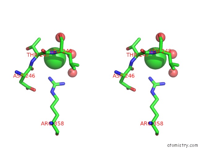
Stereo pair view
Mono view
Stereo pair view
A full contact list of Chlorine with other atoms in the Cl binding
site number 1 of Crystal Structure of N-Myristoyl Transferase (Nmt) G386E Mutant From Plasmodium Vivax within 5.0Å range:
|
Chlorine binding site 2 out of 3 in 6may
Go back to
Chlorine binding site 2 out
of 3 in the Crystal Structure of N-Myristoyl Transferase (Nmt) G386E Mutant From Plasmodium Vivax
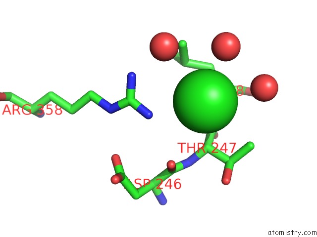
Mono view
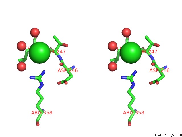
Stereo pair view
Mono view
Stereo pair view
A full contact list of Chlorine with other atoms in the Cl binding
site number 2 of Crystal Structure of N-Myristoyl Transferase (Nmt) G386E Mutant From Plasmodium Vivax within 5.0Å range:
|
Chlorine binding site 3 out of 3 in 6may
Go back to
Chlorine binding site 3 out
of 3 in the Crystal Structure of N-Myristoyl Transferase (Nmt) G386E Mutant From Plasmodium Vivax
Mono view
Stereo pair view
Mono view
Stereo pair view
A full contact list of Chlorine with other atoms in the Cl binding
site number 3 of Crystal Structure of N-Myristoyl Transferase (Nmt) G386E Mutant From Plasmodium Vivax within 5.0Å range:
|
Reference:
A.C.Schlott,
S.Mayclin,
A.R.Reers,
O.Coburn-Flynn,
A.S.Bell,
J.Green,
E.Knuepfer,
D.Charter,
R.Bonnert,
B.Campo,
J.Burrows,
S.Lyons-Abbott,
B.L.Staker,
C.W.Chung,
P.J.Myler,
D.A.Fidock,
E.W.Tate,
A.A.Holder.
Structure-Guided Identification of Resistance Breaking Antimalarial N‐Myristoyltransferase Inhibitors. Cell Chem Biol V. 26 991 2019.
ISSN: ESSN 2451-9456
PubMed: 31080074
DOI: 10.1016/J.CHEMBIOL.2019.03.015
Page generated: Sat Jul 12 16:44:34 2025
ISSN: ESSN 2451-9456
PubMed: 31080074
DOI: 10.1016/J.CHEMBIOL.2019.03.015
Last articles
Zn in 4ZZIZn in 4ZZH
Zn in 4ZYB
Zn in 4ZYO
Zn in 4ZY0
Zn in 4ZY1
Zn in 4ZYA
Zn in 4ZX9
Zn in 4ZX8
Zn in 4ZXS