Chlorine »
PDB 6n3y-6ndf »
6n47 »
Chlorine in PDB 6n47: The Structure of Sb-2-204-Tubulin Complex
Protein crystallography data
The structure of The Structure of Sb-2-204-Tubulin Complex, PDB code: 6n47
was solved by
K.Arnst,
S.Banerjee,
Y.Wang,
W.Li,
D.Miller,
W.Li,
with X-Ray Crystallography technique. A brief refinement statistics is given in the table below:
| Resolution Low / High (Å) | 50.00 / 2.60 |
| Space group | P 21 21 21 |
| Cell size a, b, c (Å), α, β, γ (°) | 105.295, 157.835, 182.068, 90.00, 90.00, 90.00 |
| R / Rfree (%) | 20.1 / 24.6 |
Other elements in 6n47:
The structure of The Structure of Sb-2-204-Tubulin Complex also contains other interesting chemical elements:
| Magnesium | (Mg) | 4 atoms |
| Calcium | (Ca) | 4 atoms |
Chlorine Binding Sites:
The binding sites of Chlorine atom in the The Structure of Sb-2-204-Tubulin Complex
(pdb code 6n47). This binding sites where shown within
5.0 Angstroms radius around Chlorine atom.
In total 3 binding sites of Chlorine where determined in the The Structure of Sb-2-204-Tubulin Complex, PDB code: 6n47:
Jump to Chlorine binding site number: 1; 2; 3;
In total 3 binding sites of Chlorine where determined in the The Structure of Sb-2-204-Tubulin Complex, PDB code: 6n47:
Jump to Chlorine binding site number: 1; 2; 3;
Chlorine binding site 1 out of 3 in 6n47
Go back to
Chlorine binding site 1 out
of 3 in the The Structure of Sb-2-204-Tubulin Complex

Mono view
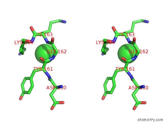
Stereo pair view
Mono view
Stereo pair view
A full contact list of Chlorine with other atoms in the Cl binding
site number 1 of The Structure of Sb-2-204-Tubulin Complex within 5.0Å range:
|
Chlorine binding site 2 out of 3 in 6n47
Go back to
Chlorine binding site 2 out
of 3 in the The Structure of Sb-2-204-Tubulin Complex
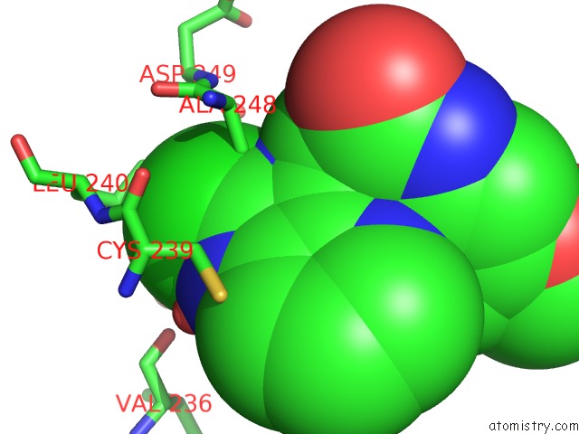
Mono view
Stereo pair view
Mono view
Stereo pair view
A full contact list of Chlorine with other atoms in the Cl binding
site number 2 of The Structure of Sb-2-204-Tubulin Complex within 5.0Å range:
|
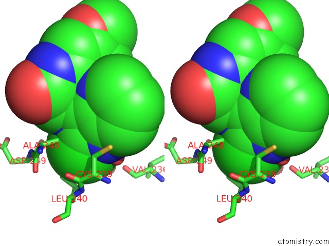
Chlorine binding site 3 out of 3 in 6n47
Go back to
Chlorine binding site 3 out
of 3 in the The Structure of Sb-2-204-Tubulin Complex

Mono view

Stereo pair view
Mono view
Stereo pair view
A full contact list of Chlorine with other atoms in the Cl binding
site number 3 of The Structure of Sb-2-204-Tubulin Complex within 5.0Å range:
|
Reference:
K.E.Arnst,
S.Banerjee,
Y.Wang,
H.Chen,
Y.Li,
L.Yang,
W.Li,
D.D.Miller,
W.Li.
X-Ray Crystal Structure Guided Discovery and Antitumor Efficacy of Dihydroquinoxalinone As Potent Tubulin Polymerization Inhibitors. Acs Chem.Biol. 2019.
ISSN: ESSN 1554-8937
PubMed: 31714738
DOI: 10.1021/ACSCHEMBIO.9B00696
Page generated: Sat Jul 12 17:01:11 2025
ISSN: ESSN 1554-8937
PubMed: 31714738
DOI: 10.1021/ACSCHEMBIO.9B00696
Last articles
I in 3WN5I in 3WYX
I in 3WGW
I in 3WD6
I in 3WB5
I in 3W31
I in 3WB4
I in 3W1N
I in 3W0F
I in 3W2Z