Chlorine »
PDB 6n3y-6ndf »
6n7f »
Chlorine in PDB 6n7f: 1.90 Angstrom Resolution Crystal Structure of Glutathione Reductase From Streptococcus Pyogenes in Complex with Fad.
Enzymatic activity of 1.90 Angstrom Resolution Crystal Structure of Glutathione Reductase From Streptococcus Pyogenes in Complex with Fad.
All present enzymatic activity of 1.90 Angstrom Resolution Crystal Structure of Glutathione Reductase From Streptococcus Pyogenes in Complex with Fad.:
1.8.1.7;
1.8.1.7;
Protein crystallography data
The structure of 1.90 Angstrom Resolution Crystal Structure of Glutathione Reductase From Streptococcus Pyogenes in Complex with Fad., PDB code: 6n7f
was solved by
G.Minasov,
L.Shuvalova,
I.G.Shabalin,
M.Grabowski,
A.Olphie,
A.Cardona-Correa,
W.F.Anderson,
K.J.F.Satchell,
A.Joachimiak,
Center Forstructural Genomics Of Infectious Diseases (Csgid),
with X-Ray Crystallography technique. A brief refinement statistics is given in the table below:
| Resolution Low / High (Å) | 29.47 / 1.90 |
| Space group | P 1 |
| Cell size a, b, c (Å), α, β, γ (°) | 76.400, 86.650, 86.670, 67.13, 74.15, 74.11 |
| R / Rfree (%) | 16.9 / 21 |
Chlorine Binding Sites:
Pages:
>>> Page 1 <<< Page 2, Binding sites: 11 - 17;Binding sites:
The binding sites of Chlorine atom in the 1.90 Angstrom Resolution Crystal Structure of Glutathione Reductase From Streptococcus Pyogenes in Complex with Fad. (pdb code 6n7f). This binding sites where shown within 5.0 Angstroms radius around Chlorine atom.In total 17 binding sites of Chlorine where determined in the 1.90 Angstrom Resolution Crystal Structure of Glutathione Reductase From Streptococcus Pyogenes in Complex with Fad., PDB code: 6n7f:
Jump to Chlorine binding site number: 1; 2; 3; 4; 5; 6; 7; 8; 9; 10;
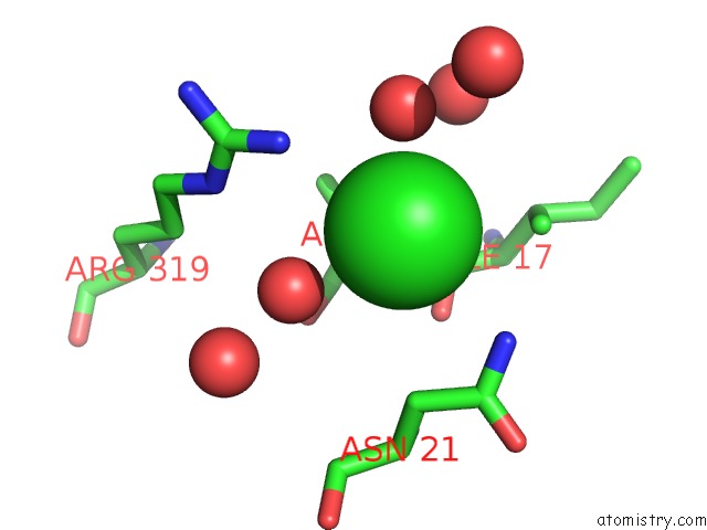
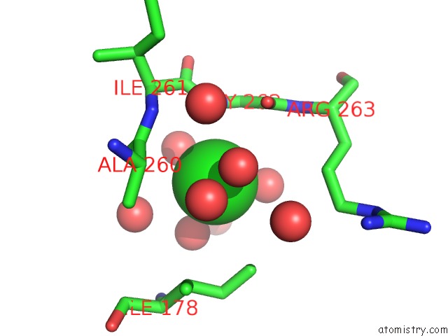
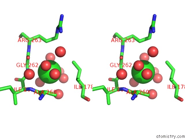
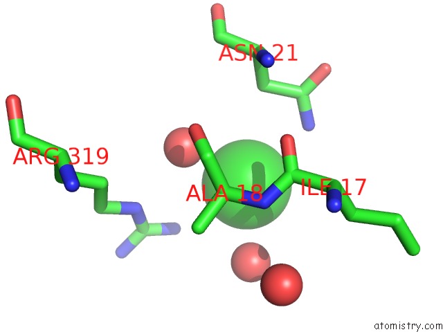
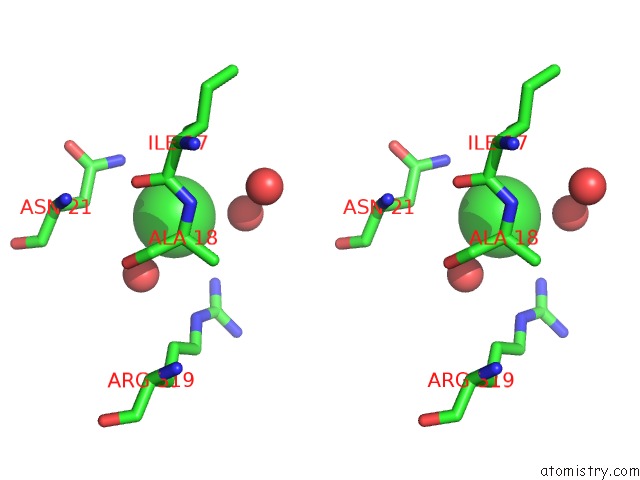
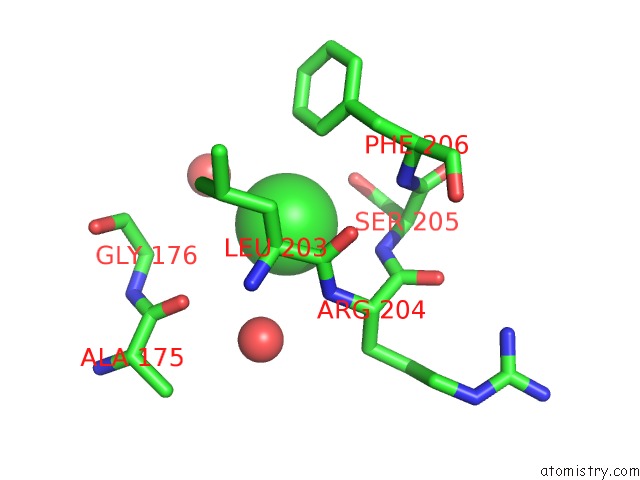
Chlorine binding site 1 out of 17 in 6n7f
Go back to
Chlorine binding site 1 out
of 17 in the 1.90 Angstrom Resolution Crystal Structure of Glutathione Reductase From Streptococcus Pyogenes in Complex with Fad.
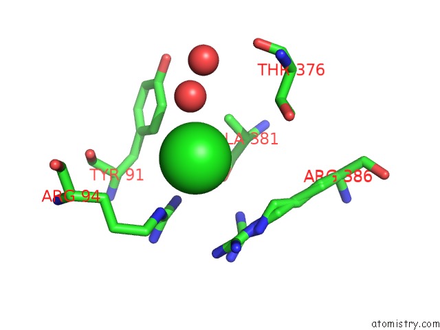
Mono view
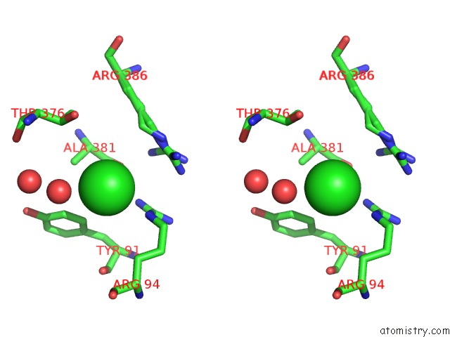
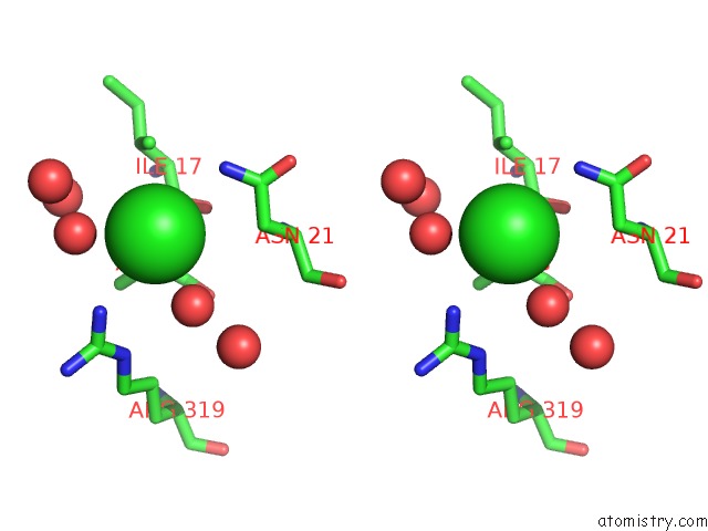
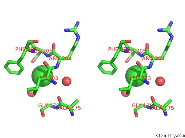
Stereo pair view
Mono view
Stereo pair view
A full contact list of Chlorine with other atoms in the Cl binding
site number 1 of 1.90 Angstrom Resolution Crystal Structure of Glutathione Reductase From Streptococcus Pyogenes in Complex with Fad. within 5.0Å range:
|
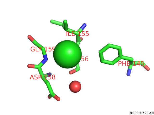
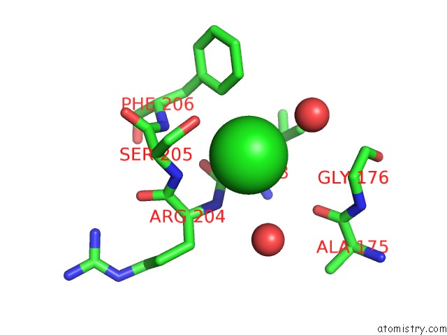
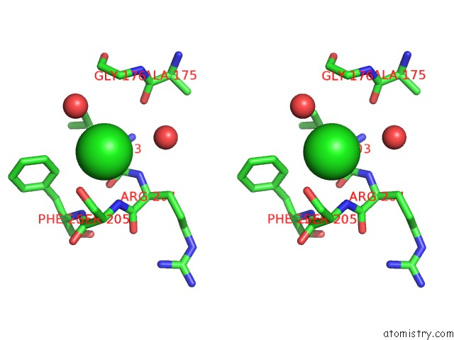
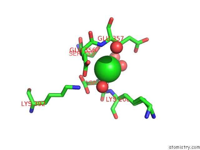
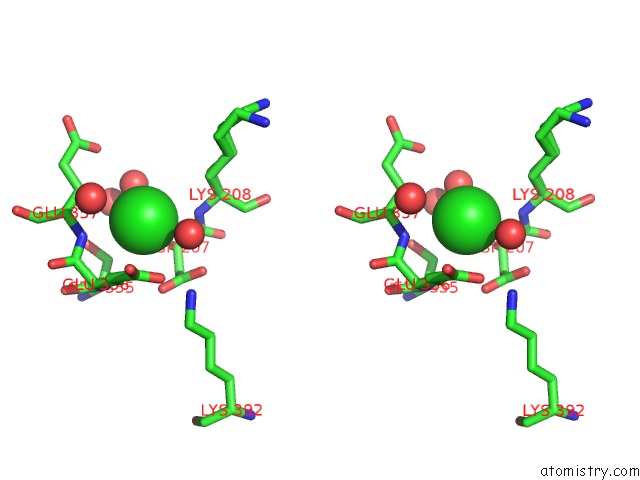
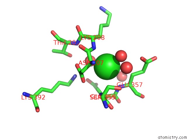
Chlorine binding site 2 out of 17 in 6n7f
Go back to
Chlorine binding site 2 out
of 17 in the 1.90 Angstrom Resolution Crystal Structure of Glutathione Reductase From Streptococcus Pyogenes in Complex with Fad.
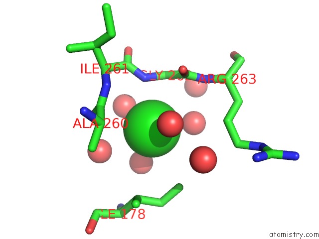
Mono view
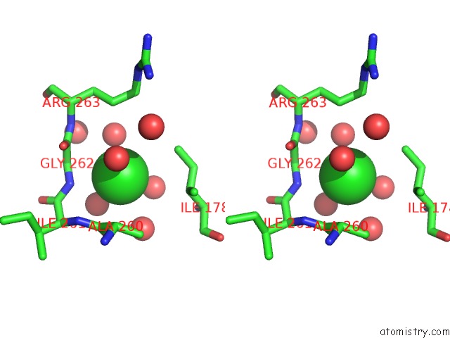
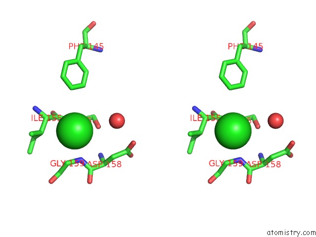
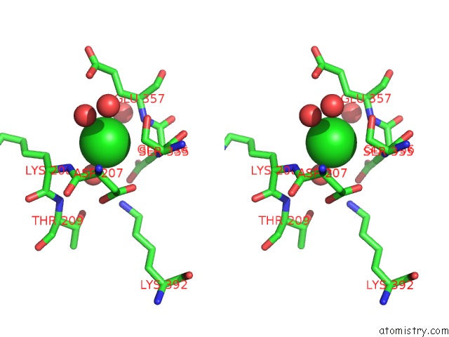
Stereo pair view
Mono view
Stereo pair view
A full contact list of Chlorine with other atoms in the Cl binding
site number 2 of 1.90 Angstrom Resolution Crystal Structure of Glutathione Reductase From Streptococcus Pyogenes in Complex with Fad. within 5.0Å range:
|
Chlorine binding site 3 out of 17 in 6n7f
Go back to
Chlorine binding site 3 out
of 17 in the 1.90 Angstrom Resolution Crystal Structure of Glutathione Reductase From Streptococcus Pyogenes in Complex with Fad.
Mono view
Stereo pair view
Mono view
Stereo pair view
A full contact list of Chlorine with other atoms in the Cl binding
site number 3 of 1.90 Angstrom Resolution Crystal Structure of Glutathione Reductase From Streptococcus Pyogenes in Complex with Fad. within 5.0Å range:
|
Chlorine binding site 4 out of 17 in 6n7f
Go back to
Chlorine binding site 4 out
of 17 in the 1.90 Angstrom Resolution Crystal Structure of Glutathione Reductase From Streptococcus Pyogenes in Complex with Fad.
Mono view
Stereo pair view
Mono view
Stereo pair view
A full contact list of Chlorine with other atoms in the Cl binding
site number 4 of 1.90 Angstrom Resolution Crystal Structure of Glutathione Reductase From Streptococcus Pyogenes in Complex with Fad. within 5.0Å range:
|
Chlorine binding site 5 out of 17 in 6n7f
Go back to
Chlorine binding site 5 out
of 17 in the 1.90 Angstrom Resolution Crystal Structure of Glutathione Reductase From Streptococcus Pyogenes in Complex with Fad.
Mono view
Stereo pair view
Mono view
Stereo pair view
A full contact list of Chlorine with other atoms in the Cl binding
site number 5 of 1.90 Angstrom Resolution Crystal Structure of Glutathione Reductase From Streptococcus Pyogenes in Complex with Fad. within 5.0Å range:
|
Chlorine binding site 6 out of 17 in 6n7f
Go back to
Chlorine binding site 6 out
of 17 in the 1.90 Angstrom Resolution Crystal Structure of Glutathione Reductase From Streptococcus Pyogenes in Complex with Fad.
Mono view
Stereo pair view
Mono view
Stereo pair view
A full contact list of Chlorine with other atoms in the Cl binding
site number 6 of 1.90 Angstrom Resolution Crystal Structure of Glutathione Reductase From Streptococcus Pyogenes in Complex with Fad. within 5.0Å range:
|
Chlorine binding site 7 out of 17 in 6n7f
Go back to
Chlorine binding site 7 out
of 17 in the 1.90 Angstrom Resolution Crystal Structure of Glutathione Reductase From Streptococcus Pyogenes in Complex with Fad.
Mono view
Stereo pair view
Mono view
Stereo pair view
A full contact list of Chlorine with other atoms in the Cl binding
site number 7 of 1.90 Angstrom Resolution Crystal Structure of Glutathione Reductase From Streptococcus Pyogenes in Complex with Fad. within 5.0Å range:
|
Chlorine binding site 8 out of 17 in 6n7f
Go back to
Chlorine binding site 8 out
of 17 in the 1.90 Angstrom Resolution Crystal Structure of Glutathione Reductase From Streptococcus Pyogenes in Complex with Fad.
Mono view
Stereo pair view
Mono view
Stereo pair view
A full contact list of Chlorine with other atoms in the Cl binding
site number 8 of 1.90 Angstrom Resolution Crystal Structure of Glutathione Reductase From Streptococcus Pyogenes in Complex with Fad. within 5.0Å range:
|
Chlorine binding site 9 out of 17 in 6n7f
Go back to
Chlorine binding site 9 out
of 17 in the 1.90 Angstrom Resolution Crystal Structure of Glutathione Reductase From Streptococcus Pyogenes in Complex with Fad.
Mono view
Stereo pair view
Mono view
Stereo pair view
A full contact list of Chlorine with other atoms in the Cl binding
site number 9 of 1.90 Angstrom Resolution Crystal Structure of Glutathione Reductase From Streptococcus Pyogenes in Complex with Fad. within 5.0Å range:
|
Chlorine binding site 10 out of 17 in 6n7f
Go back to
Chlorine binding site 10 out
of 17 in the 1.90 Angstrom Resolution Crystal Structure of Glutathione Reductase From Streptococcus Pyogenes in Complex with Fad.
Mono view
Stereo pair view
Mono view
Stereo pair view
A full contact list of Chlorine with other atoms in the Cl binding
site number 10 of 1.90 Angstrom Resolution Crystal Structure of Glutathione Reductase From Streptococcus Pyogenes in Complex with Fad. within 5.0Å range:
|
Reference:
G.Minasov,
L.Shuvalova,
I.G.Shabalin,
M.Grabowski,
A.Olphie,
A.Cardona-Correa,
W.F.Anderson,
K.J.F.Satchell,
A.Joachimiak,
Center For Structural Genomics Of Infectious Diseases(Csgid).
1.90 Angstrom Resolution Crystal Structure of Glutathione Reductase From Streptococcus Pyogenes in Complex with Fad. To Be Published.
Page generated: Sat Jul 12 17:02:17 2025
Last articles
K in 9ES1K in 9EIO
K in 9ED1
K in 9EIE
K in 9EAF
K in 9DWN
K in 9E4V
K in 9DKF
K in 9DTR
K in 9DXX